
bone scan
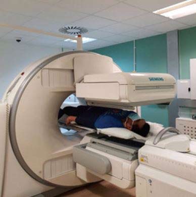
A bone scan is an imaging test that shows areas of increased or decreased bone turnover. A bone scan involves injecting a radioactive material (radiotracer) into a vein. The substance travels through the bloodstream to the bones and organs. As it wears away, it gives off radiation, which is detected by a camera that slowly scans the patient's body. The camera takes pictures of how much radiotracer collects in the bones.
If a bone scan is done to see if there is a bone infection, images may be taken shortly after the radioactive material is injected and again 3 to 4 hours later, when it has collected in the bones. This is called a 3-phase bone scan.
To evaluate metastatic bone disease (see metastasis), images are taken only after the 3 to 4 hour delay.
The scanning part of the test lasts about 1 hour. The scanner's camera may move above and around the patient, so there is no need for him or her to change positions.
Why a bone scan is performed
A bone scan is used to:
Results of a bone scan
Test results are considered normal if the radiotracer moves evenly throughout all the bones in your body. The images should show that the radioactive material has been evenly distributed throughout the body. There should be no areas of increased or decreased distribution. "Hot spots" are areas where there is an increased accumulation of the radioactive material. "Cold spots" are areas that have taken up less of the radioactive material.
Risks
If you are pregnant or nursing, the test may be postponed to prevent exposing the developing baby to radiation. If you must have the test while breast-feeding, you should pump and throw away the breast milk for the next 2 days.
The amount of radiation injected into your vein is very small, and nearly all radiation is gone from the body within 2–3 days. The radiotracer that is used exposes you to a very small amount of radiation. The risk is probably no greater than with routine or conventional X-rays. Risks related to the bone radiotracer are rare, but may include:

