
chest X-ray
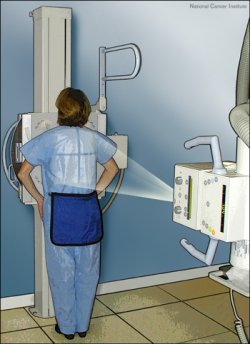
How a chest X-ray is carried out. Credit: National Cancer Institute.

Chest X-ray of normal appearance taken from the back of an adult male.
A chest X-ray is an X-ray of the chest, lungs, heart, large arteries, ribs, and diaphragm. The test is carried out in a hospital radiology department or in a health care provider's office by an X-ray technician. Two views are usually taken: one in which the X-rays pass through the chest from the back (posterior-anterior view), and one in which the X-rays pass through the chest from one side to the other (lateral view). You stand in front of the machine and must hold your breath when the X-ray is taken.
A chest X-ray may be ordered when a person's symptoms include a persistent cough, coughing up blood, chest pain, a chest injury, or difficulty in breathing. The test is also used when tuberculosis, lung cancer, or other chest or lung disease is suspected. A serial chest X-ray (repeated or sequential) may be used to evaluate changes (for example, an increase in the size of an abnormality) found on a previous chest X-ray.
Risks
There is low radiation exposure. X-rays are monitored and regulated to provide the minimum amount of radiation exposure needed to produce the image. Most experts feel that the risk is very low compared with the benefits. Pregnant women and children are more sensitive to the risks of the X-ray. Chest X-rays are generally avoided during the 1st and 2nd trimesters of pregnancy (a trimester is a period of 3 months).

