
epiglottic cartilage
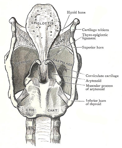
Posterior aspect of cartilages and ligaments of the larynx.
The epiglottic cartilage is a thin, leaf-like lamina of elastic fibrocartilage placed behind the tongue and the body of the body of the hyoid bone, in the anterior boundary of the inlet and vestibule of the larynx. When divested of the mucous membrane, the cartilage of the epiglottis has the form of an obovate leaf; and it shows numerous pits and perforations. Glands are lodged in the pits; and foramina transmit vessels and nerves. The broad end of the cartilage is directed upwards, and is free; its lateral margins are to a large extent enclosed within the aryepiglottic folds. The anterior surface is free only in its upper part. That part is covered with mucous membrane, and looks towards the tongue. The posterior surface is covered with mucous membrane throughout its whole extent. The lower end of the cartilage is pointed, and is connected with the thyroid cartilage by the thyroepiglottic ligament.
The epiglottis is bound by mucous folds and by ligaments to the back of the tongue, to the side wall of the pharynx, to the hyoid bone, and to the thyroid cartilage.
The hyoepiglottic ligament is a short, broad band of loose fibroelastic tissue which connects the anterior face of the epiglottic cartilage to the upper border of the body of the hyoid bone. The thyroepiglottic ligament is strong, elastic and thick, and attaches the lower end of the cartilage to the back of the thyroid cartilage a little below the median notch.
The triangular interval between the lower part of the epiglottis and the median thyrohyoid ligament contains a pad of soft fat, and it is imperfectly closed above by the hyoepiglottic ligament.

