
peptidoglycan
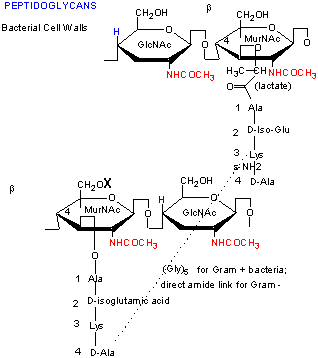
Peptidoglycan is a cross-linked complex of polysaccharides and peptides found in the cell walls of bacteria.
Peptidoglycan in the cell wall of Gram-positive bacteria
Gram-positive bacteria (so-called because they color violet when treated appropriately with Gram's stain) have a thick layer of a peptidoglycan (or murein), the form of which determines the organism's shape – bacilli (rod shaped), cocci (spherical shaped), or spirilla (helical shaped).
• Gram-positive bacteria have a large quantity of peptidoglycan in the cell wall – typically 40–60% of the dry weight of the wall.
• The wall is homogenous.
• Lipids and proteins are generally absent from the wall.
• Considerable cross-linking occurs between adjacent peptidoglycan strands
• Gram-positive bacteria have teichoic acids.
Peptidoglycan in the cell wall of Gram-negative bacteria
In contrast, gram-negative organisms have only a very thin layer of peptidoglycan immediately outside their cell membrane (about one twentieth of the thickness of that found in gram-positive organisms). Surrounding this thin wall of peptidoglycan, however, gram-negative organisms have a bilayered membrane composed of phospholipid and bacterial lipopolysaccharide which has the ability to protect the internal structures of the microbe from damaging chemicals.
• Gram-negative bacteria have a low degree of cross-linking and a low peptidoglycan content, seldom exceeding 5–10% of the wall weight.
• Peptidoglycan forms the innermost layer of a multilayered wall.
• Superimposed on the thin peptidoglycan layer is an outer cell wall layer with a structure typical of a unit membrane. The outer cell wall (outer membrane) consists of proteins, phospholipids, and lipopolysaccharides.

