
skull, muscular attachments
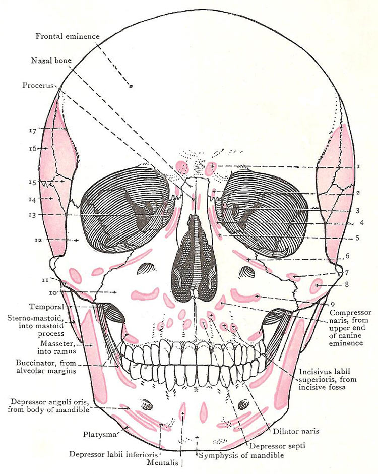
Anterior (front) view of the skull
showing the places where muscles attach.
1, 2, 3, 4. Orbicularis oculi.
5. Levator labii superioris alaeque nasi.
6. Levator labii.
7. Zygomaticus minor (from zygomatic bone).
8. Zygomaticus major (from zygomatic bone).
9. Levator anguli oris (from maxilla).
10. Zygomatic process of maxilla.
11. Masseter, from zygomatic arch.
12. Frontal process of zygomatic bone.
13. Lacrimal bone and fossa.
14. Squamous temporal (from temporal muscle).
15. Greater wing of sphenoid (from temporal muscle).
16. Parietal bone.
17. Temporal line.
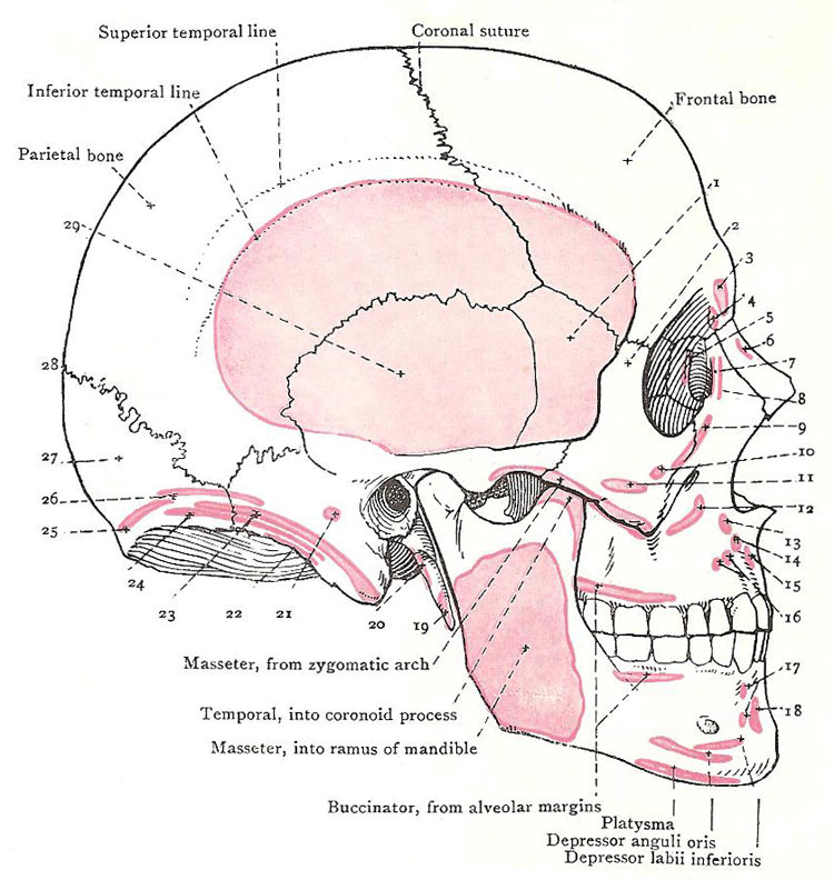
Lateral (side) view of the skull showing
the places where muscles attach.
1. Greater wing of sphenoid bone.
2. Frontal process of zygomatic bone.
3, 4. Orbicularis oculi (from frontal bone).
5. Orbicularis (from lacrimal crest).
6. Procerus and nasal bone.
7. Orbicularis (from frontal process of maxilla).
8. Levator labii superioris alaeque nasi.
9. Levator labii superioris.
10. Zygomaticus minor (from zygomatic bone).
11. Zygomaticus major (from zygomatic bone).
12. Levator anguli oris (from maxilla).
13. Compressor naris.
14. Dilator naris.
15. Depressor septi.
16, 17. Incisive muscles.
18. Mentalis (from body of mandible).
19. Styroglossus (from styloid process).
20. Styrohyoid (from styloid process).
21. Auricularis posterior (on mastoid temporal).
22. Longissimus capitis (on mastoid temporal).
23. Sternomastoid.
24. Splenius capitis (on superior nuchal line).
25. Trapezius (on superior nuchal line).
26. Occipital belly of occipito frontalis.
27. Squamous part of occipital bone.
28. Lambda.
29. Temporal muscle and squamous temporal.

