
spinal nerve

Spinal nerves pass through the intervertebral canals.

Connections of the spinal nerve roots to the spinal cord.

The branches of a spinal nerve.

Typical spinal nerve, with a cross section of the spinal cord.
A spinal nerve is any of the nerves that arise in pairs from the spinal cord. The spinal nerves are distinguished from the cranial nerves, which, apart from the vagus nerve, are limited in their distribution to the head and neck. Like the cranial nerves, the spinal nerves are part of the peripheral nervous system.
There are 31 pairs of spinal nerves in the human body: 8 cervical, 12 thoracic, 5 lumbar, 5 sacral, and 1 coccygeal. These emerge through spaces between the vertebrae. The uppermost pair passes through the intervertebral canals between the occipital bone and the atlas, while the lowest pair passes through the intervertebral canal in the coccyx.
Each spinal nerve has two nerve roots (except the first, which has no sensory root). The root in the front, called the anterior or ventral root, transmits impulses from the spinal cord to the muscles. The root in the back, known as the posterior or dorsal root, carries sensory information (about touch, position, pain, and temperature) from the body to the spinal cord.
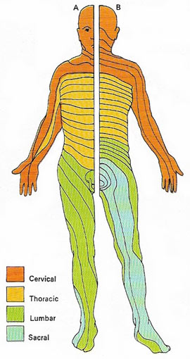 |
| Spinal nerves carrying messages to and from the spinal cord serve specific areas of skin known as dermatomes. The 31 pairs, one for each of the bones of he spine (the vertebrae), are subdivided into five groups. As the area served by the last pair of spinal nerves is ill-defined, only the four major groups are shown for the front (A) and back (B).
|
Motor and sensory roots
The anterior root of each spinal nerve is made of nerve fibers which come from the anterior column of the spinal cord. It consists solely of motor nerve fibers. The posterior root springs from the posterior part of the spinal cord, and consists of fibers which lead to the posterior column of the cord. These fibers are all sensory fibers. On the posterior there is a small bulge. It is called the posterior root ganglion and is caused by the bodies of the nerve cells which form the sensory nerve fibers.
Within the spinal canal the anterior and posterior roots unite to form a mixed spinal nerve. This nerve leaves the spinal canal through the intervertebral canal.
Branches of the spinal nerves
As each spinal nerve leaves the intervertebral canal it divides into two branches. The smaller branch curves sharply backwards and is called the posterior primary ramus. It contains both motor and sensory fibers. Soon it divides into many small branches to supply the muscles and the skin of the back.
The large branch of each spinal nerve is called the anterior primary ramus. It, too, contains both motor and sensory nerve fibers. The anterior ramus runs forward in the tissues, dividing into many small branches which serve the muscles and the skin of the front of the body.
In the lower neck region and the lumbar region the anterior rami of the spinal nerves are very large. In each of these regions they fuse together in groups in a complicated way to form nerve plexuses. From these plexuses spring the series of large nerves which supply motor nerve fibers to the muscles, and sensory nerve fibers to the skin of the arms and the legs.

