pneumothorax

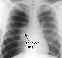
X-ray of right lung pneumothorax.

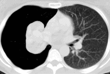
CT of right lung pneumothorax
Source: John Hopkins School of Medicine.
Pneumothorax is a condition in which air enters the pleural cavity (the space between the two layers of the pleura which cover the lungs and chest wall). The air may enter the pleural cavity from the lungs or from outside the body, expanding the normally closed pleural space, raising pleural pressure toward atmospheric pressure, and resulting in partial or complete collapse of the lung. When pleural pressure approaches zero, the lung and chest wall both move toward the equilibrium positions they would assume in the absence of any external pressures – the lung collapses and the chest wall springs out.
Causes
There are various causes of pneumothorax. Spontaneous pneumothorax, which usually occurs for no apparent reason, is six times more likely to occur in men than in women. Most often, it affects thin young adults who have no underlying lung disease; in many cases, it is thought to be due to rupture of a congenital blister at the top of the lung. There is a 30% chance of recurrence of spontaneous pneumothorax, usually on the same side.
Pneumothorax may also be a complication of lung disease, particularly asthma or emphysema.
Traumatic pneumothorax may follow an injury, such as a fractured rib. It may also be caused accidentally when a catheter is inserted into a vein in the neck for intravenous feeding or to monitor pressure in the heart and circulation.
Symptoms
A pneumothorax may cause chest pain or shortness of breath. The degree of breathlessness is proportional to the size of the pneumothorax. Any underlying lung disease will increase breathing difficulty. If there is continual leakage of air into the pleural cavity, the pneumothorax may become progressively bigger and produce a tension pneumothorax, which may become life-threatening because of compression of the heart.
Diagnosis and treatment
A chest X-ray confirms the diagnosis. A small pneumothorax in a healthy adult usually disappears within a few days without treatment. A larger pneumothorax, or a small one in the presence of underlying lung disease, requires treatment.
Treatment usually involves removing the air from the pleural cavity through a suction tube inserted through the chest wall for several days. A small pneumothorax can be treated by drawing out the air through a needle and syringe. If the lung fails to expand, or if the pneumothorax recurs, surgery may be required to seal the pleural cavity.


