
arytenoid cartilages
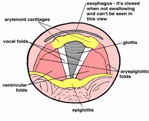
Horizontal section through the larynx, showing the vocal folds in relationship to other structures, including the arytenoid cartilages.
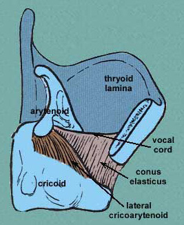
Vertical section through the larynx (Adam's apple to the right).
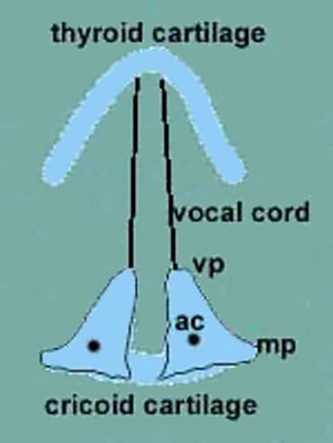
The arrangement of cartilages in the larynx. ac = arytenoid cartilage; mp = muscular process; vp = vocal process.
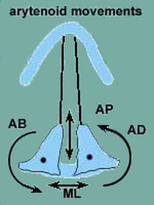
The movements that take place between the arytenoid and cricoid cartilages (cricoarytenoid joints). The dot in the arytenoid cartilage is the vertical axis around which the arytenoid cartilage rotates.
The arytenoid cartilages are a pair of small cartilages, shaped roughly like three-sided pyramids, which are located toward the rear of the larynx and play a vital role in positioning and adjusting the vocal folds when speaking or singing.
The base of each arytenoid cartilage is broad and has a smooth surface for articulation with the cricoid cartilage (the lowest cartilage in the larynx). From its base, sitting atop the back edge of the cricoid, each arytenoid cartilage points forward. The foremost point of the base of each arytenoid cartilage is called the vocal process, and it is to this point that the rear of each vocal fold is attached. The vocal folds then run forward to insert at a point at the inside center of the thyroid cartilage. The pointed tip, or apex, of each arytenoid cartilage is covered by a small nodule called the corniculate cartilage.
The arytenoid cartilages are connected to small muscles by which they can move the vocal folds, enabling them to become tensed, to various degrees, and relaxed. When vocalizing, the cartilages need to be in close proximity to hold the vocal folds together.
Arytenoid movements
The relationship of the arytenoid cartilages to the cricoid and thyroid cartilages is shown in the upper illustration below. The ways in which the arytenoid cartilages can move are illustrated in the second of the two illustrations below. These movements include:
adduction (AD)
abduction (AB)
anterior-posterior sliding (AP)
medial-lateral sliding (ML)
To understand how each of these movements is carried out and the muscles involve, see:
transverse
arytenoid muscle
lateral cricoarytenoid
muscles
thyroarytenoideus muscle
posterior cricoarytenoid
muscle
See also abduction and adduction of vocal folds.

