
alveolus
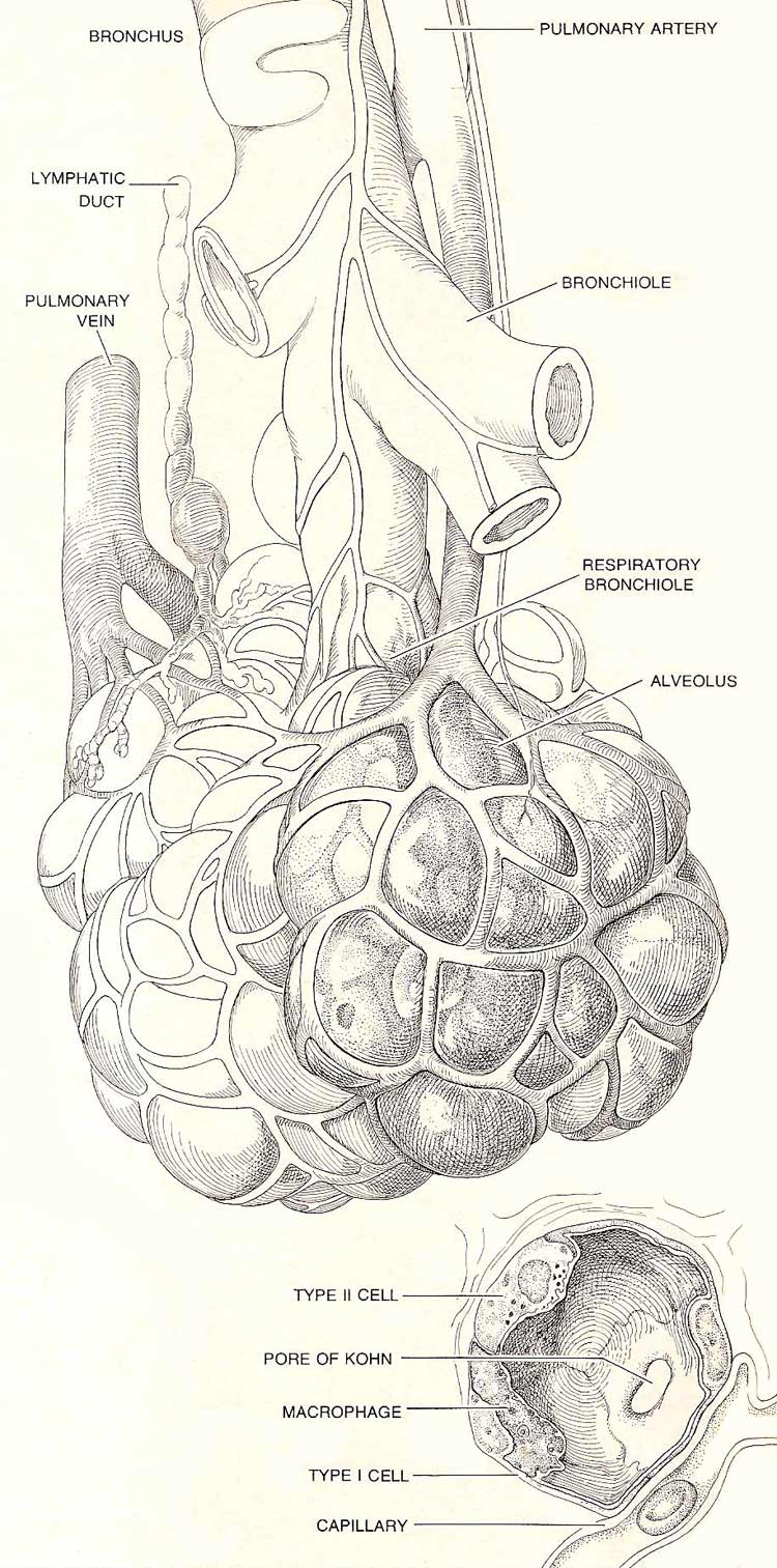
Figure 1. Clusters of alveoli, forming alveolar sacs, are connected by ducts to the smallest air pathways of the lung: the respiratory bronchioles. Several sacs and the network of capillaries that surrounds them are illustrated here. Seen in cross section is a single alveolus with its lining of three distinctive kinds of cell. Cells labeled Type II secrete lipids that reduce surface tension during exhalation and keep the lung from deflating completely.
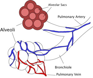
Figure 2. Simplified diagram of alveoli.
1. An alveolus is a minute air-filled sac in the vertebrate lung (Figures 1 and 2). Pulmonary alveoli are thin-walled and surrounded by blood vessels. There are large numbers of alveoli in each lung, and it is through their surfaces that the respiratory exchange of oxygen and carbon dioxide occurs. In most vertebrates they connect with the mouth by a system of ramifying air-tubes – the bronchi and bronchioles.
Alveolitis is a condition in which the walls of the alveoli become inflamed and thickened, causing the lungs to become less elastic and less able to transfer oxygen.
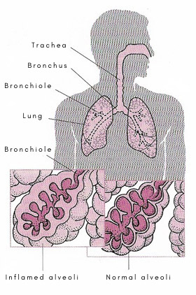 |
| Effects of alveolitis |
2. The term alveolus may also mean an expanded sac of secretory epithelium, which forms from an internal termination of each duct in many glands (e.g. the mammary glands).
3. A dental alveolus is a cavity in the jaw-bone into which a tooth fits.

