
choroid
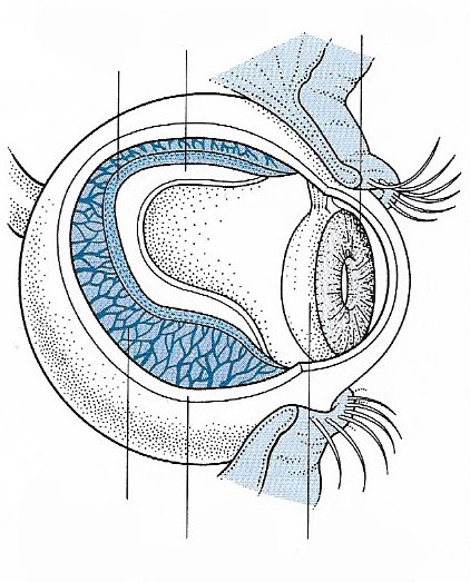
Figure 1. The choroid lines the inside of the eyeball. It thickens around the lens to form the ciliary body. Muscles stretching between the choroid and the lens contract to adjust the shapeof the lens in focusing.

Figure 2. The three main layers of the vertebrate eye: retina, choroid, and sclera. Image credit: University of Texas Medical Branch.
The choroid is a layer in the vertebrate eye, which lies immediately
outside the retina, between the retina and
the sclera. The choroid contains blood vessels,
which carry nourishment to the outer layers of the retina, and pigment.
The choroid connects with the ciliary body toward the front of the eye and is attached to edges of the optic
nerve at the back of the eye.
The choroid itself consists of four layers: an outermost layer of large
blood vessels (Haller's layer), a layer of intermediate
size blood vessels (Sattler's layer), a layer of capillaries (choriocapillary layer), and an innermost layer (Bruch's
membrane).
In primates, including humans, dark-colored melanin in the choroid helps restrict the amount of internal reflection in the eye, which might otherwise result in the perception of multiple, confusing images. Poor vision in albinos stems from a lack of this pigmentation. By contrast, the choroid of many other animals contains reflective materials that help to collect light in dim situations.
The "red eye" seen on photos, especially those taken with a flash, is caused by reflection of light by the choroid. The redness is due to the choroid's numerous blood vessels.

