
femoral artery
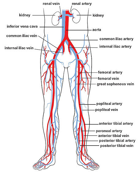
Major arteries and veins of the lower body.
The femoral artery is a major blood vessel that supplies oxygenated blood to the leg. The femoral artery is the direct continuation of the external iliac artery of the abdomen, and is the great arterial trunk of the leg. It begins behind the inguinal ligament, at the mid-inguinal point, and it descends through the upper two-thirds of the thigh to the opening in the adductor magnus, through which it passes into the popliteal artery. The extent of the artery in the femoral triangle and in the subsartorial canal varies with the width of the sartorius, but usually the upper half is in the triangle and the lower half in the canal.
Relations of the femoral artery
The relations which the artery bears to the femur are important. As it enters the thigh it leaves the brim of the pelvis and lies in front of the medial part of the head of the femur, from both of which it is separated by the psoas major muscle. During the remainder of its course through the femoral triangle, the artery is not in close relation to the bone; it crosses in front of the angular interval between the neck and shaft of the femur. Toward the apex of the triangle, however, it comes into relation with the medial surface of the shaft of the femur, being separated only by the posterior part of the vastus medialis, which is thin there; that position it holds to its termination, but, owing to the obliquity of the medial surface of the femur, the terminal part of the artery is well behind the bone.
In the femoral triangle, the artery is quite superficial, being covered only by skin and fasciae and crossed at the apex of the triangle by the medial cutaneous nerve. The only structure related to the artery medially is the femoral vein, in the upper part of the triangle; but it takes up a position behind the artery in the lower part of the triangle. Lateral to the artery are the femoral nerve and its branches.
Behind the artery are the muscles of the floor of the triangle. But it is separated from the pectineus by a mass of fat which contains the profunda vessels, and from the adductor longus by the femoral vein. At apex of the triangle, the order of structures, from front to back, is: femoral artery, femoral vein, adductor longus, profunda vein, and profunda artery; in a stab-wound at that point all four vessels may be severed.
In the subsartorial canal, the artery is separated from the adductor longus and magnus by the femoral vein, which is posterior to the artery in the upper part of the canal and posterolateral in the lower part; and the adductor longus separates the femoral vessels from the profunda vessels. The nerve to the vastus medialis is lateral to the artery in the upper part of the canal; and the saphenous nerve crosses gradually in front of the artery from lateral to medial side.
Branches of the femoral artery
In addition to the three small superficial arteries of the groin and the deep external pudendal, the artery gives off, in the femoral triangle, the profunda femoris. The branches which arise in the subsartorial canal are muscular twigs and the descending genicular artery, which springs from the femoral trunk a short distance above the opening in the adductor magnus. It gives branches to muscles and to the knee joint, and one accompanies the saphenous nerve to the medial side of the knee and the leg.

