A

David

Darling

melanoma

melanoma

Melanoma.


melanomas

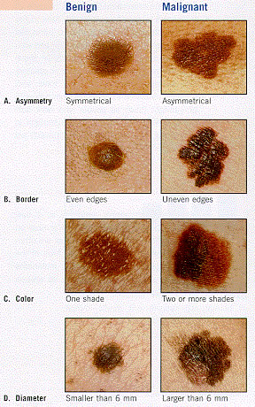
The ABCD rule for spotting a melanoma.


Melanoma is a form of skin cancer that begins in melanocytes – the cells that make the brown pigment melanin. Melanoma usually starts in a mole.


The skin is the body's largest organ. It protects against heat, sunlight, injury, and infection. The skin has two main layers: the epidermis (upper or outer layer) and the dermis (lower or inner layer).


Melanocytes are found throughout the lower part of the epidermis. They make melanin, the pigment that gives skin its natural color. When skin is exposed to the sun, melanocytes make more pigment, causing the skin to tan, or darken.


When melanoma starts in the skin, the disease is called cutaneous melanoma. Melanoma may also occur in the eye and is called intraocular or ocular melanoma.


Melanoma is the most aggressive of three types of skin cancer, the others being basal cell skin cancer and squamous cell skin cancer.


Where on the body does melanoma occur?

Melanoma can occur anywhere on the body.
In men, melanoma is often found on the trunk (the area from the shoulders to the hips) or the head and neck. In women, melanoma often develops on the arms and legs. Melanoma usually occurs in adults, but it is sometimes found in children and adolescents.


What increases the risk of melanoma?

Unusual moles, exposure to sunlight, and health history can affect the risk of developing melanoma.


Anything that increases your risk of getting a disease is called a risk factor. Risk factors for melanoma include the following:


  • Unusual moles
  • Exposure to natural sunlight
  • Exposure to artificial ultraviolet light (tanning booth)
  • Family or personal history of melanoma
  • Being white and older than 20 years
  • Red or blond hair
  • White or light-colored skin and freckles
  • Blue eyes
  •  


    Signs and symptoms

    Possible signs of melanoma include a change in the appearance of a mole or pigmented area.


    These and other symptoms may be caused by melanoma or by other conditions. A doctor should be consulted if any of the following problems occur:


  • A mole that:
          - changes in size, shape, or color
          - has irregular edges or borders
          - is more than 1 color
          - is asymmetrical (if the mole is divided in half, the 2 halves are different in size or shape)
          - itches
          - oozes, bleeds, or is ulcerated (a hole forms in the skin when the top layer of cells breaks down and the underlying tissue shows through).

  • Change in pigmented (colored) skin.
  • Satellite moles (new moles that grow near an existing mole).
  •  

    Here is the "ABCD rule" to help tell a normal mole from a melanoma:


  • A: asymmetry – one half of the mole does not match the other half.
  • B: border irregularity – the edges of the mole are ragged or notched.
  • C: color – the color of the mole is not the same all over. There may be shades of tan, brown, or black, and sometimes patches of red, blue, or white.
  • D: diameter – the mole is wider than about 1/4 inch (although doctors are now finding more melanomas that are smaller).
  •  

    Other important signs of melanoma include changes in size, shape, or color of a mole. Some melanomas do not fit the descriptions above, and it may be hard to tell if the mole is normal or not, so you should show your doctor anything that you are unsure of.


    Diagnosis

    Tests that examine the skin are used to detect (find) and diagnose melanoma.


    If a mole or pigmented area of the skin changes or looks abnormal, the following tests and procedures can help detect and diagnose melanoma:


  • Skin examination: A doctor or nurse examines the skin to look for moles, birthmarks, or other pigmented areas that look abnormal in color, size, shape, or texture.

  • Biopsy: A local excision is done to remove as much of the suspicious mole or lesion as possible. A pathologist then looks at the tissue under a microscope to check for cancer cells. Because melanoma can be hard to diagnose, patients should consider having their biopsy sample checked by a second pathologist.
  •  

    Suspicious areas should not be shaved off or cauterized (destroyed with a hot instrument, an electrical current, or a caustic substance).


    What affects the chance of recovery?

    Certain factors affect prognosis (chance of recovery) and treatment options.


    The prognosis (chance of recovery) and treatment options depend on the following:


  • The stage of melanoma (whether cancer is found in the outer layer of skin only, or has spread to the lymph nodes, or to other places in the body.
  • Whether there was bleeding or ulceration at the primary site.
  • The location and size of the tumor.
  • The patient's general health.
  •  

    Although many people are successfully treated, melanoma can recur (come back). In recurrent melanoma, the cancer may come back at the original site or in other parts of the body, such as the lungs or liver.


    Stages of melanoma


    Stage 0

    In stage 0, melanoma is found only in the epidermis (outer layer of the skin). Stage 0 is also called melanoma in situ.


    Stage I

    Stage I is divided into stages IA and IB.


  • Stage IA: In stage IA, the tumor is not more than 1 millimeter thick, with no ulceration. The tumor is in the epidermis and upper layer of the dermis.

  • Stage IB: In stage IB, the tumor is either:
          - not more than 1 millimeter thick, with ulceration, and may have spread into the dermis or the tissues below the skin;
    or
          -1 to 2 millimeters thick, with no ulceration.
  •  


    Stage II

    Stage II is divided into stages IIA, IIB, and IIC.


  • Stage IIA: In stage IIA, the tumor is either:
          - 1 to 2 millimeters thick, with ulceration; or
          - 2 to 4 millimeters thick, with no ulceration.

  • Stage IIB: In stage IIB, the tumor is either:
          - 2 to 4 millimeters thick, with ulceration; or
          - more than 4 millimeters thick, with no ulceration.

  • Stage IIC: In stage IIC, the tumor is more than 4 millimeters thick, with ulceration.
  •  


    Stage III


    In stage III, the tumor may be any thickness, with or without ulceration, and:


  • has spread to 1 or more lymph nodes; or
  • has spread into the nearby lymph system but not into nearby lymph nodes; or
  • has spread to lymph nodes that are matted (not moveable); or
  • satellite tumors (additional tumor growths within 2 centimeters of the original tumor) are present and nearby lymph nodes are involved.
  •  


    Stage IV

    In stage IV, the tumor may be any thickness, with or without ulceration, may have spread to 1 or more nearby lymph nodes, and has spread to other places in the body.


    Tests done to diagnose the stage of melanoma

    The process used to find out whether cancer has spread within the skin or to other parts of the body is called staging. The information gathered from the staging process determines the stage of the disease. It is important to know the stage in order to plan treatment.


    The following tests and procedures may be used in the staging process:


  • Wide local excision: A surgical procedure to remove some of the normal tissue surrounding the area where melanoma was found, to check for cancer cells.

  • Lymph node mapping and sentinel lymph node biopsy: Procedures in which a radioactive substance and/or blue dye is injected near the tumor. The substance or dye flows through lymph ducts to the sentinel node or nodes (the first lymph node or nodes where cancer cells are likely to have spread). The surgeon removes only the nodes with the radioactive substance or dye. A pathologist then checks the sentinel lymph nodes for cancer cells. If no cancer cells are detected, it may not be necessary to remove additional nodes.

  • Chest X-ray: An X-ray of the organs and bones inside the chest. An X-ray is a type of energy beam that can go through the body and onto film, making a picture of areas inside the body.

  • CT scan (CAT scan): A procedure that makes a series of detailed pictures of areas inside the body, taken from different angles. The pictures are made by a computer linked to an X-ray machine. A dye may be injected into a vein or swallowed to help the organs or tissues show up more clearly. This procedure is also called computed tomography, computerized tomography, or computerized axial tomography. For melanoma, pictures may be taken of the chest, abdomen, and pelvis.

  • MRI (magnetic resonance imaging): A procedure that uses a magnet, radio waves, and a computer to make a series of detailed pictures of areas inside the body. This procedure is also called nuclear magnetic resonance imaging (NMRI).

  • PET scan (positron emission tomography scan): A procedure to find malignant tumor cells in the body. A small amount of radioactive glucose (sugar) is injected into a vein. The PET scanner rotates around the body and makes a picture of where glucose is being used in the body. Malignant tumor cells show up brighter in the picture because they are more active and take up more glucose than normal cells do.

  • Laboratory tests: Medical procedures that test samples of tissue, blood, urine, or other substances in the body. These tests help to diagnose disease, plan and check treatment, or monitor the disease over time.
  •  

    The results of these tests are viewed together with the results of the tumor biopsy to determine the melanoma stage.