
vestibular folds
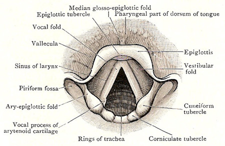
Cavity of larynx, as seen with a laryngoscope during quiet breathing – rima glottidis widely opened. Vestibular and vocal folds can be seen.
Each of the vestibular folds is a prominent fold of mucous membrane that extends, antero-posteriorly, across the side wall of the laryngeal cavity (see larynx). Each vestibular fold is soft and rather flaccid, and its free border is slightly arched, with the concavity looking downward. It contains: (1) the vestibular ligament, a weak band of fibro-elastic tissue; (2) numerous mucous glands, which are aggregated chiefly in its middle part; and (3) a few muscle fibers.
The space between the vestibular folds is called the rima vestibuli, and is wider than that between the vocal folds. It follows, therefore, that both pairs of folds are visible when the cavity of the larynx is looked at from above.

