
vocal folds
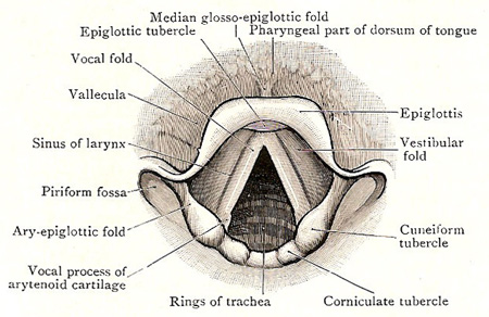
Figure 1. Cavity of larynx, as seen with a laryngoscope during quiet breathing – rima glottidis widely openedk.
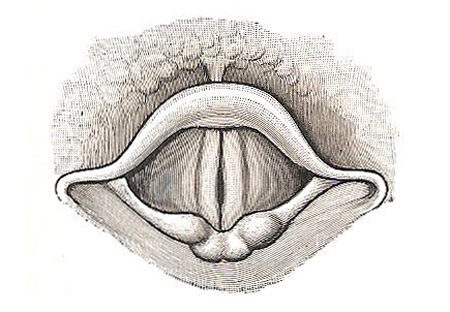
Figure 2. Cavity of larynx, as seen with a laryngoscope during phonation – rima glottidis closed.
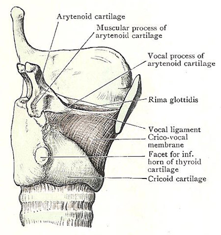
Figure 3. Larynx, side view, showing vocal ligament.
The vocal folds are structures in the larynx by means of which the voice is produced (Figures 2 and 3). Each vocal fold is sharp and prominent, and its mucous membrane is thin and firmly bound down to the vocal ligament, which is enclosed within it. In color it is pale – almost pearly white – whilst posteriorly, where the fold ends, the point of the vocal process of the arytenoid cartilage stands out in relief and has a yellowish tinge. In coronal section, the vocal fold is prismatic in form, and the free border looks upward and medially.
By contrast with the vocal folds, the vestibular folds are of little importance in producing the voice; in fact, they can be destroyed, in large part, without any appreciable effect upon the voice.
Vocal ligaments
The vocal ligament is not an isolated band, but is the thickened anterior part f the upper border of the cricovocal membrane; and it is composed almost entirely of elastic tissue (Figure 3). It lies within the vocal fold, to which it gives support; and it is closely bound to the mucous membrane of the fold. Its posterior end is attached to the tip of the vocal process of the arytenoid cartilage; the anterior part is attached to the deep surface of the thyroid cartilage opposite the middle of the anterior border, close to its fellow of the other side.

