
intercostal muscles
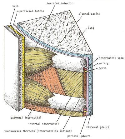
Section through an intercostal space.
The intercostal muscles are three muscles located in the intercostal space between ribs. These muscles are comparable to those of the anterior abdominal wall.
The external intercostal muscle forms the most superficial layer. Its fibers are directed downward and forward from the inferior border of the rib above to the superior border of the rib below. The muscle extends forward from the rib tubercle behind to the costochondral junction in front, where the muscle is replaced by an aponeurosis, the anterior (external) intercostal membrane.
The internal intercostal muscle forms the intermediate layer. Its fibers are directed downward and backward from the subcostal groove of the rib above to the upper border of the rib below. The muscle extends backward from the sternum in front to the angles of the ribs behind, where the muscle is replaced by an aponeurosis, the posterior (internal) intercostal membrane.
The transversus thoracis muscle forms the deepest layer and corresponds to the transversus abdominis muscle in the anterior abdominal wall. It is an incomplete muscle layer and crosses more than one intercostal space within the ribs. It is related internally to fascia (endothoracic fascia) and parallel pleura and externally to the intercostal nerves and vessels. The transversus thoracis muscle may be divided into three portions, which are more or less separate from one another; (1) the subcostalis, (2) the intercostalis intimus, and (3) the sternocostalis.
Action of intercostal muscles
When the intercostal muscles contract, they all tend to pull the ribs nearer to one another. If the first rib is fixed by the contraction of the muscles in the root of the neck, namely, the scaleni muscles, the intercostal muscles will raise the second to the twelfth ribs toward the first. If, on the other hand, the twelfth rib is fixed by the quadratus lumborum muscle and the oblique muscles of the abdomen, the first to the eleventh ribs will be lowered by the contraction of the intercostal muscles. In addition, the tone of the intercostal muscles during the different phases of respiration serves to strengthen the tissues of the intercostal space, thus preventing the sucking in or the blowing out of the tissues with changes in the intrathoracic pressure.
Nerve supply of intercostal muscles
The intercostal muscles are supplied by the corresponding intercostal nerves. The intercostal nerves and blood vessels (the neurovascular bundle), as in the abdominal wall, run between the middle and innermost layers of muscles. They are arranged in the following order from above downward: intercostal vein, intercostal artery, and intercostal nerve.

