
brain tumor
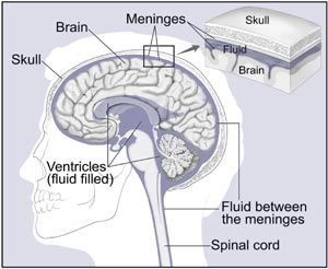
The brain and nearby structures.
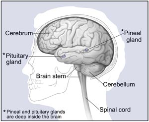
Major parts of the brain.
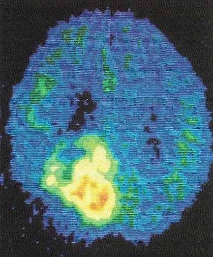
PET scan of a brain shows the presence, extent, and location of a tumor. The irregular bright area on the scan is a result of faster glocose metabolism in that part of the brain.
A brain tumor is a tumor that begins in the brain is called a primary brain tumor. In children, most brain tumors are primary tumors. In adults, most tumors in the brain have spread there from the lung, breast, or other parts of the body. When this happens, the disease is not brain cancer. The tumor in the brain is a secondary tumor. It is named for the organ or the tissue in which it began.
Treatment for secondary brain tumors depends on where the cancer started and the extent of the disease.
The brain
The brain is a soft, spongy mass of tissue. It is protected by the bones of the skull and three thin membranes called meninges. Watery fluid called cerebrospinal fluid cushions the brain. This fluid flows through spaces between the meninges and through spaces within the brain called ventricles.
A network of nerves carries messages back and forth between the brain and the rest of the body. Some nerves go directly from the brain to the eyes, ears, and other parts of the head. Other nerves run through the spinal cord to connect the brain with the other parts of the body. Within the brain and spinal cord, glial cells surround nerve cells and hold them in place.
The brain directs the things we choose to do (like walking and talking) and the things our body does without thinking (like breathing). The brain is also in charge of our senses (sight, hearing, touch, taste, and smell), memory, emotions, and personality.
The three major parts of the brain control different activities:
The cerebrum is divided into the left and right cerebral hemispheres, which control separate activities. The right hemisphere controls the muscles on the left side of the body. The left hemisphere controls the muscles on the right side of the body.
Benign and malignant brain tumors
Brain tumors can be benign or malignant:
Usually, benign tumors can be removed, and they seldom grow back.
The border or edge of a benign brain tumor can be clearly seen. Cells from benign tumors do not invade tissues around them or spread to other parts of the body. However, benign tumors can press on sensitive areas of the brain and cause serious health problems.
Unlike benign tumors in most other parts of the body, benign brain tumors are sometimes life threatening.
Very rarely, a benign brain tumor may become malignant.
Malignant brain tumors are generally more serious and often are life threatening.
They are likely to grow rapidly and crowd or invade the surrounding healthy brain tissue.
Very rarely, cancer cells may break away from a malignant brain tumor and spread to other parts of the brain, to the spinal cord, or even to other parts of the body. The spread of cancer is called metastasis.
Sometimes, a malignant tumor does not extend into healthy tissue. The tumor may be contained within a layer of tissue. Or the bones of the skull or another structure in the head may confine it. This kind of tumor is called encapsulated.
Tumor grade
Doctors sometimes group brain tumors by grade – from low grade (grade I) to high grade (grade IV). The grade of a tumor refers to the way the cells look under a microscope. Cells from high-grade tumors look more abnormal and generally grow faster than cells from low-grade tumors.
Primary brain tumors
Tumors that begin in brain tissue are known as primary tumors of the brain. (Information about secondary brain tumors appears in the following section.) Primary brain tumors are named according to the type of cells or the part of the brain in which they begin.
The most common primary brain tumors are gliomas. They begin in glial cells. There are many types of gliomas:
Some types of brain tumors do not begin in glial cells. The most common of these are:
Secondary brain tumors
When cancer spreads from its original place to another part of the body, the new tumor has the same kind of abnormal cells and the same name as the primary tumor. Cancer that spreads to the brain from another part of the body is different from a primary brain tumor. When cancer cells spread to the brain from another organ (such as the lung or breast), doctors may call the tumor in the brain a secondary tumor or metastatic tumor. Secondary tumors in the brain are far more common than primary brain tumors.
Who is at risk from brain tumors?
No one knows the exact causes of brain tumors. Doctors can seldom explain why one person develops a brain tumor and another does not. However, it is clear that brain tumors are not contagious. No one can "catch" the disease from another person.
Research has shown that people with certain risk factors are more likely than others to develop a brain tumor. A risk factor is anything that increases a person's chance of developing a disease.
The following risk factors are associated with an increased chance of developing a primary brain tumor:
Radiation. Workers in the nuclear industry have an increased risk of developing a brain tumor.
Formaldehyde. Pathologists and embalmers who work with formaldehyde have an increased risk of developing brain cancer. Scientists have not found an increased risk of brain cancer among other types of workers exposed to formaldehyde.
Vinyl chloride. Workers who make plastics may be exposed to vinyl chloride. This chemical may increase the risk of brain tumors.
Acrylonitrile. People who make textiles and plastics may be exposed to acrylonitrile. This exposure may increase the risk of brain cancer.
Scientists are investigating whether cell phones may cause brain tumors. Studies thus far have not found an increased risk of brain tumors among people who use cell phones.
Scientists also continue to study whether head injuries are a risk factor for brain tumors. So far, these studies have not found an increased risk among people who have had head injuries.
Most people who have known risk factors do not get brain cancer. On the other hand, many who do get the disease have none of these risk factors. People who think they may be at risk should discuss this concern with their doctor. The doctor may be able to suggest ways to reduce the risk and can plan an appropriate schedule for checkups.
Symptoms
The symptoms of brain tumors depend on tumor size, type, and location. Symptoms may be caused when a tumor presses on a nerve or damages a certain area of the brain. They also may be caused when the brain swells or fluid builds up within the skull.
These are the most common symptoms of brain tumors:
These symptoms are not sure signs of a brain tumor. Other conditions also could cause these problems. Anyone with these symptoms should see a doctor as soon as possible. Only a doctor can diagnose and treat the problem.
Diagnosis
If a person has symptoms that suggest a brain tumor, the doctor may perform one or more of the following procedures:
The doctor may ask for other tests:
Surgeons can obtain tissue to look for tumor cells in three ways:
Sometimes a biopsy is not possible. If the tumor is in the brain stem or certain other areas, the surgeon may not be able to remove tissue from the tumor without damaging normal brain tissue. The doctor uses MRI, CT, or other imaging tests instead.
A person who needs a biopsy may want to ask the doctor the following questions:

