
brain

Figure 1. Human brain, lateral view.
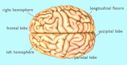
Figure 2. Human brain, seen from above.
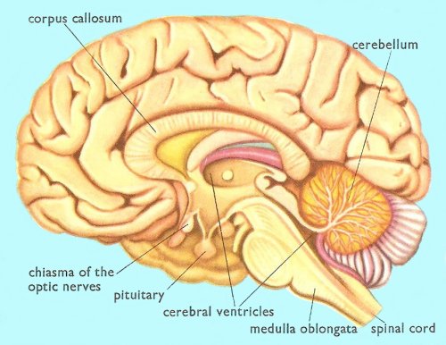
Figure 3. Human brain, lateral cross-section.
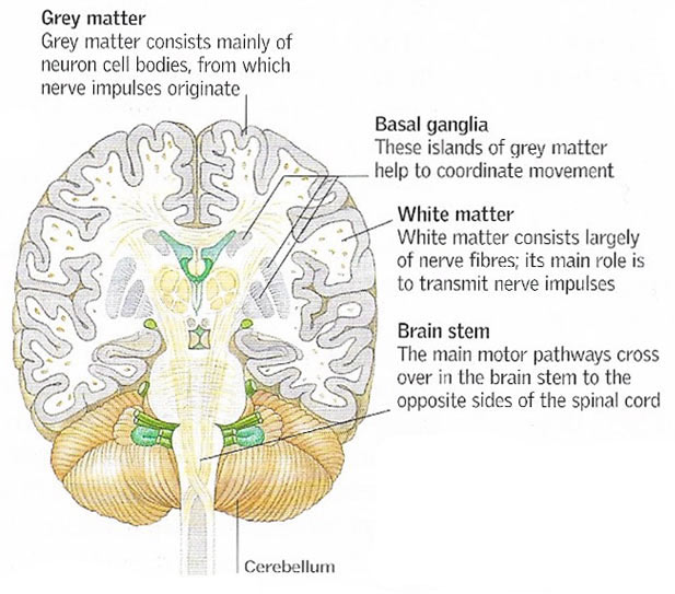
Figure 4. Brain tissue. The outer layer of the brain, the cerebral cortex, is made of gray matter. Beneath are white matter and islands of gray matter.
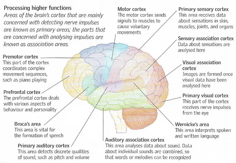
Figure 5. Different types of sensory information are processed in different parts of the nervous system, which sends out appropriate response signals. Complex information, such as data about music and emotion, is processed in the cerebral cortex.
In humans and other vertebrates, the brain is the enlarged anterior portion of the central nervous system. It is encased within the cranium of the skull and is continuous with the spinal cord. It is surrounded by three membranes, called meninges, and is bathed in cerebrospinal fluid, which fills the internal cavities, known as ventricles.
The brain serves as the main coordinating center for nervous activity, receiving information in the form of nerve impulses from sensory organs, interpreting it, and transmitting signals to muscles and other effectors. It is also the seat of intelligence and memory.
Brain development
The embryonic vertebrate brain consists of three sections, known as the forebrain, midbrain, and hindbrain, which become further differentiated during development into specialized regions. Principal among these regions in the adult are the cerebrum, the cerebellum, the medulla oblongata, and the hypothalamus.
Cerebrum
The cerebrum forms the largest part of the human brain. It is very soft and oval-shaped. The outer part of the cerebrum, called the cerebral cortex, is made up of gray matter, which consists of many layers of nerve cells. One part of the cortex is occupied by the cells that initiate movements of the voluntary muscles; another contains the cells in which the electrical nerve messages from the sense organs all over the body are converted into conscious sensations. These and other areas of the cortex are named after the functions that they perform. Thus there are motor area, sensory areas, visual areas, and auditory areas.
The cortex is crossed by winding furrows of varying depth. These are called sulci (Latin sulcus, a furrow or groove). The deeper ones divide the brain up into several parts, called lobes.
Beneath the cortex lies white matter, consisting of vast numbers of nerve fibers which help to connect the cortical cells with the sensory organs and muscles all over the body.
See from above, it is clear that the brain is divided by a deep cleft (the longitudinal fissure) into two parts known as the right and left cerebral hemispheres.
Motor areas
When a human brain is viewed from the side, one of its most conspicuous features is the deep groove which runs downwards and forwards from the top towards the temporal lobe. This groove is called the fissure of Rolando. Both in front of and behind this fissure are unusually large ridges, or gyri, of brain tissue.
The ridge in front of the fissure is often called the pre-central gyrus. It is of extreme importance because its cortex contains the nerve cells which control the movements of our voluntary muscles. Because this part of the brain makes our muscles move it is often called the motor area.
The nerve cells in this area area arranged in a manner which seems rather strange. Firstly, the motor area on each side of the brain contains the nerve cells which control the movements of muscles on the opposite side of the body. This is because the nerve fibers from the motor cortex cross to the opposite side as they pass down the spinal cord.
The second odd feature of the motor area is that the cells which control the muscles of the toes and feet lie at the top of the area while those that control the upper parts of our bodies lie at the bottom.
Those parts of the body which make rapid and accurate movements, such as the fingers and the tongue, have many cells to control their muscles and for this reason relatively large parts of the cortex are devoted to them. Those muscles which make coarse movements, even though they may be very large, are controlled by fewer cells.
Sensory areas
The gyrus which lies immediately behind the fissure of Rolando is known as the post-central gyrus. This part of the brain is connected to the nerves which carry messages from the sense organs in our skin and muscles. Here are registered the sensations of touch, pressure, heat, and cold. This is the reason why the post-central gyrus is often called the sensory area.
As in the case of the motor areas, each sensory area serves the opposite side of the body. Furthermore, the various parts of the body are again represented upside-down on the surface of the gyrus. As a result of this, the motor and sensory areas for each part of the body are close together, one on either side of the fissure.
The sensory functions of the brain are not confined to the post-central gyrus, but they extend backwards over several nearby areas of the cortex. These neighboring areas seem to be concerned with the more subtle of our sensory functions. They play an important part in our ability to distinguish between small differences in the weight, temperature, and texture of objects. In addition they are concerned with the faculty of stereognosis, or the ability to identify, without seeing them, small objects that are held in the hands.
Special senses
Sight, hearing, taste, and smell are known as the special senses. The degree to which each is developed varies greatly in different animal species. Humans, for example, have eyesight which is excellent when compared with that of many animals, but the human sense of smell is very poor compared with that of the dog.
Each of the special sense organs – the eyes, the ears, the olfactory organ, and the taste buds in the mouth – is connected with the brain by a cranial nerve. These nerves lead to particular areas of the cortex which are devoted to the reception of the sensations provided by the organs of the special senses. The optic nerves from the eyes carry their electrical messages to the cortex of the occipital lobe at the very hindmost part of the brain. It is here that the visual sensations are appreciated.
The auditory nerves from the ears join the brain on its under-surface. They run a complicated course inside the brain tissue before they finally reach the cortex of the upper part of the temporal lobes.
The nerves which carry the sensations of taste end in the post-central cortex close to the area devoted to the mouth and tongue. Although it often seems that this part of our nervous system is designed merely to add to the pleasures of eating, there is reason to believe that the sense of taste is important in the selection of those types of food which the body needs at any particular time.
Speech and writing
These very complicated skills are among the most sophisticated of which the human brain is capable. Both have to be learned, and in consequence they are largely dependent on the senses of hearing and sight. The sounds of speech are carried to the auditory area, as are all sounds, but it is in the surrounding area of the temporal lobe, sometimes called the auditory speech center, that recognition of their meaning takes place. In a similar way the visual images of letters and words are carried to the visual area in the cortex of the occipital lobes, but their meanings are understood in the adjacent area.
In right-handed people, the sensory centers for both spoken and written words are on the left side of the brain. Both of them are connected by many nerve fibers to a part of the left prefrontal lobe known as Broca's area after the man who first tried to locate it. The functions of this area may be to plan the muscular actions which are required in speech and in writing, and then to send appropriate instructions to those parts of the motor cortex which control the muscles of the larynx and the right hand. The impulses generated in the cells of the motor cortex pass along the motor nerves to cause the vocal cords to move in speech and the fingers to move as the pen forms letters on the page.
Prefrontal lobes
Those parts of the brain in the front of the skull just above the eyes are called the prefrontal lobes. The cortex in this area if os particular importance in humans because it is involved in the processes of learning, thinking, and reasoning. It seems that part of what we call human intelligence depends on the proper functioning of this area of the brain.
In addition to intelligence, the prefrontal lobes are concerned with the manner in which individuals behave. They seem to be able to control the way in which a person responds to his surroundings and by so doing they determine our personalities and characters.
Cerebellum
The cerebellum is a body about 8 to 10 centimeters wide by 6 centimeters long and 5 centimeters thick, which lies under the occipital lobes. It is made up of two hemispheres and a small central part called the vermis (Latin vermis, worm) because of its appearance.
Other components
Various separate structures are found in the lower part of the brain, underneath the cerebrum. These include:
Comparison of brains
The average adult human brain has a mass of about 3 pounds (1,300-1,400 grams), or 2% of the total body mass. By contrast, an elephant's brain weighs about 6,000 grams and a cat's brain about 30 grams.
In invertebrates, the brain, where present at all, is a concentration of nerve cells usually located at the anterior end of the animal.
| Human brain: Statistics |
|---|
| The brain contains about 100 billion neurons. Neurons multiply at a rate of 250,000 neurons/minute during early pregnancy. The total surface area of the cerebral cortex is about 2500 sq. cm (~2.5 ft2). |

