
choroid plexus
The choroid plexus is a rich network of blood vessels located in the brain, which is responsible for the production of cerebrospinal fluid (CSF), the fluid which surrounds and cushions the brain and spinal cord. The choroid plexus and the arachnoid membrane act together at the barriers between the blood and CSF (see blood-brain barrier).
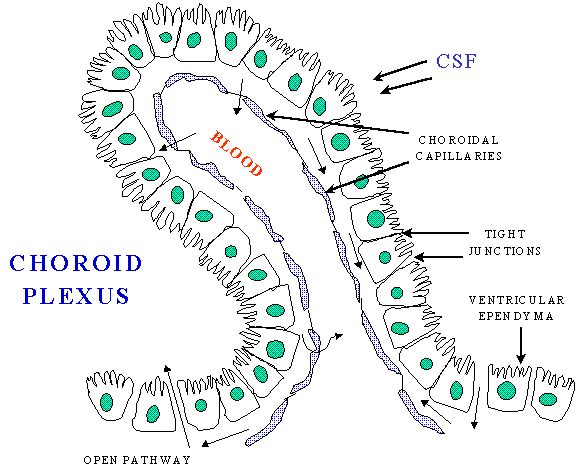
The choroid plexus not only forms the CSF but also actively regulates the concentration of molecules in it. The choroid plexus consists of highly vascularized, cauliflower-like masses of pia mater tissue that dip into pockets formed by ependymal cells. The preponderance of choroid plexus is distributed throughout the fourth ventricle near the base of the brain and in the lateral ventricles inside the right and left hemispheres of the cerebrum. The cells of the choroidal epithelium are modified and have epithelial characteristics. These ependymal cells have microvilli on the CSF side, basolateral interdigitations, and abundant mitochondria. The ependymal cells, which line the ventricles, form a continuous sheet around the choroid plexus. While the capillaries of the choroid plexus are fenestrated, non-continuous and have gaps between the capillary endothelial cells allowing the free-movement of small molecules, the adjacent choroidal epithelial cells form tight junctions preventing most macromolecules from effectively passing into the CSF from the blood. However, these epithelial-like cells have shown a low resistance as compared with the cerebral endothelial cells, between blood and CSF.

