heart electrical system
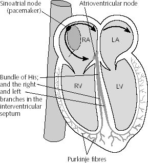
Diagram of heart chambers showing spread of excitation and sites of heart block. (RA, LA: right and left atrium. RV, LV: right and left ventricle.)
Each electrical signal in the heart begins in a group of cells called the sinus node or sinoatrial (SA) node. The SA node is located in the right atrium, which is the upper right chamber of the heart. In a healthy adult heart at rest, the SA node fires off an electrical signal to begin a new heartbeat 60 to 100 times a minute.
From the SA node, the signal travels to the right and left atria. This causes the atria to contract and pump blood into the heart's two lower chambers, the ventricles. This is recorded as the P wave on an EKG.
The signal passes between the atria and ventricles through a group of cells called the atrioventricular (AV) node. The signal slows down as it passes through the AV node. This slowing allows the ventricles time to finish filling with blood. On an EKG, this is the flat line between the end of the P wave and beginning of the Q wave.
The electrical signal then leaves the AV node and travels along a pathway called the bundle of His. From there the signal travels into the right and left bundle branches. On an EKG, this is the Q wave.
As the signal spreads across the right and left ventricles, they contract and pump blood out to the lungs and the rest of the body. On an EKG, R marks the contraction of the left ventricle and S marks the contraction of the right ventricle.
The ventricles then relax (shown as the T wave on an EKG). This entire process continues over and over with each new heartbeat.