
electrocardiogram
An electrocardiogram, also called an EKG or ECG, is a simple, non-invasive test that detects and records the electrical activity of the heart. It is used to detect and locate the source of heart problems.
Electrical signals in the heart trigger heartbeats (see heart electrical system). These signals start at the top of the heart in an area called the right atrium. The electrical signals travel from the top of the heart to the bottom. They cause the heart muscle to contract as they travel through the heart. As the heart contracts, it pumps blood out to the rest of the body.
An EKG shows how fast the heart is beating. It shows the heart's rhythm (steady or irregular) and where in the body the heartbeat is being recorded. It also records the strength and timing of the electrical signals as they pass through each part of the heart.
An EKG is sometimes called a 12-lead EKG (or 12-lead ECG) because the electrical activity of the heart is most often recorded from 12 different places on the body at the same time.
What an EKG reveals
Many heart problems change the electrical signature of the heart in distinct ways. EKG recordings of this electrical activity can help reveal a number of heart problems, including:
EKG recordings can help doctors diagnose a heart attack that is happening now or has happened in the past. This is especially true if doctors can compare a current EKG recording to an older one. EKG recordings can also reveal:
An EKG also reveals whether the heartbeat starts at the top right part of the heart like it should. It shows how long it takes for the electrical signals to travel through the heart.
Why is an electrocardiogram done?
An electrocardiogram (EKG) is done to evaluate signs and symptoms that could indicate heart problems. Some of the signs and symptoms that might be evaluated with an EKG include:
When an adult – usually someone who is older than 40 or 50 years of age – has a routine health exam, the doctor may order an EKG to screen for early heart disease that has no symptoms. The doctor is more likely to look for early heart disease if the person has a family history of heart disease in a mother, father, brother, or sister – especially if the heart disease developed early in those family members' lives.
Doctors also use EKGs to check how well heart treatments, such as drugs or medical devices, are working.
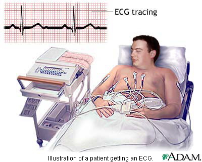
During an electrocardiogram
An electrocardiogram (EKG) is painless and harmless. A technician first attaches 12 soft patches called electrodes to the skin of the chest, arms, and legs. These electrodes are about the size of a quarter. To help an electrode stick to the skin, the technician may have to shave a patch of hair where the electrode will be attached.
After the electrodes are placed on the skin, the patient lies still on a table for a few minutes while the electrodes detect the electrical signals of the heart. A machine then records these signals on graph paper or displays them on a screen.
The entire test takes about 10 minutes. After the test, the electrodes are removed from the skin and discarded.
Special EKGs
The common EKG described above, called a resting 12-lead EKG, records only a few minutes of heart signals at a time. It will show a heart problem only if the problem is present during the few minutes that the test is being run. Many heart problems are present all the time and will be found by a resting 12-lead EKG. But some heart problems, like those related to irregular heartbeat, can come and go. They may be present for only a few minutes out of the day or only while exercising.
Special types of EKGs are used to help diagnose those kinds of problems. Three of these special EKGs are:
Stress test
Some heart problems are easier to diagnose when your heart is working harder and beating faster than when it's at rest. During stress testing, you exercise (or are given medicine if you are unable to exercise) to make your heart work harder and beat faster while heart tests are performed.
During exercise stress testing, your blood pressure and EKG readings are monitored while you walk or run on a treadmill or pedal a bicycle. Other heart tests, such as nuclear heart scanning or echocardiography, also can be done at the same time. These would be ordered if your doctor needs more information than the exercise stress test can provide about how well your heart is working.
If you are unable to exercise, a medicine can be injected through an intravenous line (IV) into your bloodstream to make your heart work harder and beat faster, as if you are exercising on a treadmill or bicycle. Nuclear heart scanning or echocardiography is then usually done.
During nuclear heart scanning, radioactive tracer is injected into your bloodstream, and a special camera shows the flow of blood through your heart and arteries. Echocardiography uses sound waves to show blood flow through the chambers and valves of your heart and to show the strength of your heart muscle.
Your doctor also may order two newer tests along with stress testing if more information is needed about how well your heart works. These new tests are magnetic resonance imaging (MRI) and positron emission tomography (PET) scanning of the heart. MRI shows detailed images of the structures and beating of your heart, which may help your doctor better assess if parts of your heart are weak or damaged. PET scanning shows the level of chemical activity in different areas of your heart. This can help your doctor determine if enough blood is flowing to the areas of your heart. A PET scan can show decreased blood flow caused by disease or damaged muscles that may not be detected by other scanning methods.

