
arrhythmias
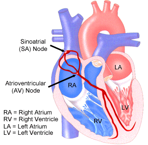
The SA and AV nodes in the heart.
An arrhythmia is an irregularity with the speed or rhythm of the heartbeat.
During an arrhythmia, the heart can beat too
fast, too slow, or with an irregular rhythm. A heartbeat that is too fast
is called tachycardia. A heartbeat that is too slow is
called bradycardia.
Atrial fibrillation
Atrial flutter
Paroxysmal supraventricular tachycardia
Ventricular tachycardia
Ventricular fibrillation
Heart attack
Heart failure or cardiomyopathy, which weakens the heart and changes
the way electrical signals move around the heart
Heart tissue that is too thick or stiff or that hasn't formed normally
Leaking or narrowed heart valves,
which make the heart work too hard and can lead to heart failure
Congenital problems (problems that are present at birth) with the
heart's structure or function
High blood pressure
Infections that damage the heart muscle or the sac around the heart
Diabetes, which increases
the risk of high blood pressure and coronary artery disease
Sleep apnea (when breathing becomes
shallow or stops during sleep), which can stress the heart because it
doesn't get enough oxygen
Overactive or underactive thyroid gland (too much or too little thyroid
hormone in the body)
Palpitations (a feeling that your heart has skipped a beat or is beating
too hard)
A slow heartbeat
An irregular heartbeat
Feeling of pauses between heartbeats
Anxiety
Weakness
Dizziness and light-headedness
Fainting or nearly fainting
Sweating
Shortness of breath
Chest pain
Cardiologists (doctors who take care of adults with heart problems)
Pediatric cardiologists (doctors who take care of babies and children
with heart problems)
Electrophysiologists (cardiologists or pediatric cardiologists who
specialize in arrhythmias)
Symptoms. What symptoms are you having? Is there a feeling of fluttering
in your chest? Do you feel dizzy or light-headed?
Medical history, including other health problems, such as a history
of heart disease, high blood pressure, diabetes, or thyroid problems.
Family medical history. Does anyone in your family have a history
of arrhythmias? Has anyone in your family ever had heart disease or
high blood pressure? Has anyone died suddenly? Are there other illnesses
or health problems in your family?
Medicines you're taking, including over-the-counter medicines and
vitamin or mineral or nutritional supplements.
Health habits, such as physical activity, smoking, or using alcohol
or drugs (for example, cocaine).
Check your pulse to find out how fast your heart is beating
Check for swelling in your legs or feet, which could be a sign of
an enlarged heart or heart failure
Look for signs of other diseases (such as thyroid disease) that could
be causing the problem
Holter monitor. This device
records the heart's electrical activity continuously over a 24-hour
period.
Event monitor. Event monitors
are useful to diagnose arrhythmias that only occur once in a while.
The device is worn continuously, but only records the heart's electrical
activity when you push a button on the device. You push the button on
the device when you feel symptoms. Event monitors can be worn for 1
to 2 months, or as long as it takes to get a recording of the heart
during symptoms.
Blood tests. These tests check
the level of substances in the blood, such as potassium or thyroid hormone,
that can increase your chances of having an arrhythmia.
Chest X-ray. A chest X-ray takes
a picture of your heart and lungs. It can show whether the heart is
enlarged.
Echocardiogram. This test uses
sound waves to create a moving picture of your heart. Echocardiogram
provides information about the size and shape of your heart and how
well your heart chambers and valves are functioning. The test also can
identify areas of poor blood flow to the heart, areas of heart muscle
that are not contracting normally, and previous injury to the heart
muscle caused by poor blood flow.
There are several different types of echocardiograms, including a stress echocardiogram. During this test, an echocardiogram is done both before and after your heart is stressed either by having you exercise or by injecting a medicine into your bloodstream that makes your heart beat faster and work harder. A stress echocardiogram is usually done to find out if you have decreased blood flow to your heart (coronary artery disease).
Transesophageal echocardiography, or TEE. This is a special type of
echocardiogram that takes pictures of the back of the heart through
the esophagus (the tube leading from your mouth to your stomach).
Stress test. Some heart problems
are easier to diagnose when your heart is working harder and beating
faster than when it's at rest. During stress testing, you exercise (or
are given medicine if you are unable to exercise) to make your heart
work harder and beat faster while heart tests are performed.
During exercise stress testing, your blood pressure and EKG readings are monitored while you walk or run on a treadmill or pedal a bicycle. Other heart tests, such as nuclear heart scanning or echocardiography, also can be done at the same time. These would be ordered if your doctor needs more information than the exercise stress test can provide about how well your heart is working.
If you are unable to exercise, a medicine can be injected through an intravenous line (IV) into your bloodstream to make your heart work harder and beat faster, as if you are exercising on a treadmill or bicycle. Nuclear heart scanning or echocardiography is then usually done.
During nuclear heart scanning, radioactive tracer is injected into your bloodstream, and a special camera shows the flow of blood through your heart and arteries. Echocardiography uses sound waves to show blood flow through the chambers and valves of your heart and to show the strength of your heart muscle.
Electrophysiologic study (EPS). This test is used to assess serious
arrhythmias. During an EPS, a thin, flexible wire is passed through
a vein in your groin (upper thigh) or arm up to the heart. The wire
records the heart's electrical signals. Your doctor uses the wire to
electrically stimulate your heart and trigger an arrhythmia. This allows
the doctor to see whether an antiarrhythmia medicine can stop the problem.
Radio-frequency ablation, a procedure used to fix some types of arrhythmia,
may be done during an EPS.
Tilt table testing. This test is sometimes used to help determine
the cause of fainting spells. You lie on a table that moves from a lying
down to an upright position. The change in position can bring on loss
of consciousness. The doctor monitors your symptoms, heart rate, EKG,
and blood pressure throughout the test. The doctor also may give you
a medicine and then monitor your response to the medicine.
Coronary angiography.
This test is an X-ray exam of the heart and blood vessels. The doctor
passes a catheter (thin, flexible tube)
through an artery in your leg or arm up to the heart. The catheter measures
the pressure inside the heart and blood vessels. A dye that can be seen
on X-ray is injected into the blood through the tip of the catheter.
The dye lets the doctor study the flow of blood through the heart and
blood vessels, which helps to diagnose blockages that can cause a heart
attack.
Gagging
Holding your breath and bearing down (Valsalva maneuver)
Immersing your face in ice-cold water
Coughing
Putting your fingers on your eyelids and pressing down gently
Keep all your medical appointments. Always bring all medicines you're
taking to all of your doctor visits. This helps ensure that all of your
doctors know exactly what medicines you're taking, which can help prevent
medication errors.
Follow your doctor's instructions for taking medicines. Check with
your doctor before taking over-the-counter medicines, nutritional supplements,
or cold and allergy medicines.
Tell your doctor if you are having side effects from your medicines.
side effects could include depression and palpitations. These side effects
can often be treated.
Tell your doctor if arrhythmia symptoms are getting worse or if you
have new symptoms.
Allow your doctor to monitor you regularly if you're taking blood-thinning
medicines.
Most arrhythmias are harmless, but some can be serious or even life threatening. When the heart rate is too slow, too fast, or irregular, the heart may not be able to pump enough blood to the body. Lack of blood flow can damage the brain, heart, and other organs.
Understanding the heart's electrical system
The heart has an internal electrical system that controls the speed and rhythm of the heartbeat. With each heartbeat, an electrical signal spreads from the top of the heart to the bottom. As it travels, the electrical signal causes the heart to contract and pump blood. The process repeats with each new heartbeat.
Each electrical signal begins in a group of cells called the sinus node, or sinoatrial (SA) node. The SA node is located in the right atrium, which is the upper right chamber of the heart. In a healthy adult heart at rest, the SA node fires off an electrical signal to begin a new heartbeat 60 to 100 times a minute.
From the SA node, the electrical signal travels through special pathways to the right and left atria. This causes the atria to contract and pump blood into the heart's two lower chambers, the ventricles. The electrical signal then moves down to a group of cells called the atrioventricular (AV) node, located between the atria and the ventricles. Here, the signal slows down just a little, allowing the ventricles time to finish filling with blood. The electrical signal then leaves the AV node and travels along a pathway called the bundle of His. This pathway divides into a right bundle branch and a left bundle branch. The signal goes down these branches to the ventricles, causing them to contract and pump blood out to the lungs and the rest of the body. The ventricles then relax, and the heartbeat process starts all over again in the SA node.
A problem with any part of this process can cause an arrhythmia. For example, in atrial fibrillation, a common type of arrhythmia, electrical signals travel through the atria in a fast and disorganized way. This causes the atria to quiver instead of contract.
Overview
There are many different types of arrhythmia. Most arrhythmias are harmless but some are not. The outlook for a person with an arrhythmia depends on the type and severity of the arrhythmia. Even serious arrhythmias can often be successfully treated. Most people with arrhythmias are able to live normal, healthy lives.
Types of arrhythmia
There are four main types of arrhythmia: premature (extra) beats, supraventricular arrhythmias, ventricular arrhythmias, and bradyarrhythmias.
Premature (extra) beats
Premature beats are the most common type of arrhythmia. They are harmless most of the time and often don't cause any symptoms. When symptoms do occur, they usually feel like a fluttering in the chest or a sensation of a skipped beat. Most of the time, premature beats need no treatment, especially in healthy people.
Premature beats that occur in the atria are called premature atrial contractions, or PACs. Premature beats that occur in the ventricles are called premature ventricular contractions, or PVCs.
In most cases, premature beats occur naturally, not due to any heart disease. But certain heart diseases can cause premature beats. They also can happen because of stress, too much exercise, or too much caffeine or nicotine.
Supraventricular arrhythmias
Supraventricular arrhythmias are tachycardias (fast heart rates) that start in the atria or the atrioventricular node (cells located between the atria and the ventricles). Types of supraventricular arrhythmias include atrial fibrillation (AF), atrial flutter, paroxysmal supraventricular tachycardia (PSVT), and Wolff-Parkinson-White (WPW) syndrome.
Atrial fibrillation
AF is the most common type of serious arrhythmia. It's a very fast and irregular contraction of the atria. AF occurs when the heart's electrical signal begins in a different part of the atrium than the sinoatrial (SA) node or when the signal is conducted abnormally. When this happens, the electrical signal doesn't travel through the normal pathways in the atria, but instead may spread throughout the atria in a fast and disorganized manner. This causes the walls of the atria to quiver very fast (fibrillate) instead of beating normally. As a result, the atria aren't able to pump blood into the ventricles the way they should.
In AF, electrical signals can travel through the atria at a rate of more than 300 per minute. Some of these abnormal electrical signals can travel to the ventricles, causing them to beat too fast and with an irregular rhythm. AF is not usually life threatening, although it can be dangerous when it causes the ventricles to beat very fast.
The two most serious complications of chronic (long-term) AF are stroke and heart failure. Stroke can happen when a blood clot travels to an artery in the brain, blocking off blood flow. In AF, blood clots can form in the atria because some of the blood pools in the fibrillating atria instead of flowing into the ventricles. If a piece of a blood clot in the left atrium breaks off, it can travel to the brain, causing a stroke. People with AF are often treated with blood-thinning medicines to reduce the chances of developing blood clots.
Heart failure is when the heart can't pump enough blood to meet the needs of the body. AF can cause heart failure when the ventricles beat too fast and don't have enough time to fill with blood to pump out to the body. Heart failure causes tiredness, leg swelling, and shortness of breath.
AF and other supraventricular arrhythmias can occur for no apparent reason. Most of the time, however, they are caused by an underlying condition that damages the heart muscle and its ability to conduct electrical impulses. These conditions include high blood pressure (hypertension), coronary artery disease, heart failure, or rheumatic heart disease.
Other conditions also can lead to AF, including overactive thyroid gland (too much thyroid hormone produced) and heavy alcohol use. AF also becomes more common as people get older.
Atrial flutter
Atrial flutter is similar to atrial fibrillation, but instead of the electrical signals spreading through the atria in a fast and irregular rhythm, they travel in a fast and regular rhythm. Atrial flutter is much less common than atrial fibrillation, but has similar symptoms and complications.
Paroxysmal supraventricular tachycardia
PSVT is a very fast heart rate that begins and ends suddenly. PSVT occurs due to problems with the electrical connection between the atria and the ventricles. In PSVT, electrical signals that begin in the atria and travel to the ventricles can reenter the atria, causing extra heartbeats. This type of arrhythmia is not usually dangerous and tends to occur in young people. It can happen during vigorous exercise.
A special type of PSVT is called Wolff-Parkinson-White syndrome. WPW syndrome is a condition in which the heart's electrical signals travel along an extra pathway from the atria to the ventricles. This extra pathway disrupts the timing of the heart's electrical signals and can cause the ventricles to beat very fast. This type of arrhythmia can be life threatening.
Ventricular arrhythmias
These are arrhythmias that start in the ventricles. They can be very dangerous and usually need immediate medical attention. Ventricular arrhythmias include ventricular tachycardia and ventricular fibrillation (v-fib). Coronary artery disease, heart attack, weakened heart muscle, and other problems can cause ventricular arrhythmias.
Ventricular tachycardia
Ventricular tachycardia is a fast, regular beating of the ventricles that may last for only a few seconds or for much longer. A few beats of ventricular tachycardia often don't cause problems, but ventricular tachycardia episodes that last for more than just a few seconds can be dangerous. Ventricular tachycardia can turn into other, more dangerous arrhythmias, such as v-fib.
Ventricular fibrillation
V-fib occurs when disorganized electrical signals make the ventricles quiver instead of pump normally. Without the ventricles pumping blood out to the body, a person will lose consciousness within seconds and will die within minutes if not treated. To prevent death, the condition must be treated immediately with defibrillation, an electric shock to the heart. V-fib may happen during or after a heart attack, or in a heart that is already weak because of another condition. Health experts think that most of the sudden cardiac deaths that occur every year (about 335,000) are due to v-fib.
Torsades de pointes (torsades) is a specific form of v-fib with a unique pattern on an EKG (electrocardiogram). Certain medicines and imbalanced amounts of potassium, calcium, or magnesium in the bloodstream can cause this condition. People with a particular finding on an EKG test, called prolonged QT interval, are at increased risk of developing torsades. People with prolonged QT interval need to be careful about taking certain antibiotics, heart medicines, and over-the-counter medicines.
Bradyarrhythmias
Bradyarrhythmias are arrhythmias in which the heart rate is slower than normal. If the heart rate is too slow, not enough blood reaches the brain, and the person can lose consciousness. In adults, a heart rate slower than 60 beats per minute is considered a bradyarrhythmia. Some people normally have slow heart rates, especially people who are very physically fit. For them, a heartbeat slower than 60 beats per minute is not dangerous and doesn't cause symptoms. But in other people, bradyarrhythmia can be due to a serious disease or other condition.
Bradyarrhythmias can be caused by heart attack, conditions that harm or change the heart's electrical system (such as underactive thyroid gland or aging), an imbalance of chemicals or other substances (such as potassium) in the blood, or even some medicines (such as beta blockers).
Bradyarrhythmias also can happen as a result of severe bundle branch block. Bundle branch block is a condition in which the electrical signal traveling down either or both of the bundle branches is delayed or blocked. When this happens, the ventricles don't contract at exactly the same time, as they should, and the heart has to work harder to pump blood to the body. The cause of bundle branch block is often an existing heart condition.
Arrhythmias in children
Normally, a child's heart beats between 70 and 100 times a minute. A newborn's heart beats about 140 times a minute. A baby or child's heart can beat faster or slower than normal for many reasons. As is true for adults, when children are active, their hearts will beat faster. When they are sleeping, their heart will beats slower. Their heart rates can speed up and slow down as they breathe in and out. All of these changes are normal.
Some children are born with heart defects that cause arrhythmias. In other children, arrhythmias can develop later in childhood. Doctors do the same kinds of tests in children and adults to diagnose arrhythmias.
Treatments for children with arrhythmias include medicines, electric shock (defibrillation), surgically implanted devices that control the heartbeat, and other procedures that fix distorted electrical signals in the heart.
What causes an arrhythmia?
An arrhythmia can occur when the electrical signals that control the heartbeat are delayed or blocked. This can happen when the special nerve cells that produce the electrical signal don't work properly or when the electrical signal doesn't travel normally through the heart. An arrhythmia also can occur when another part of the heart starts to produce electrical signals, adding to the signals from the special nerve cells and disrupting the normal heartbeat.
Stress, smoking, heavy alcohol use, heavy exercise, use of certain drugs (such as cocaine or amphetamines), use of certain prescription or over-the-counter medicines, and too much caffeine or nicotine can lead to arrhythmia in some people.
A heart attack or an underlying condition that damages the heart's electrical system also can cause an arrhythmia. These conditions include high blood pressure (hypertension), coronary artery disease, heart failure, overactive or underactive thyroid gland (too much or too little thyroid hormone produced), and rheumatic heart disease.
For some arrhythmias, such as Wolff-Parkinson-White syndrome, the underlying heart defect that causes the arrhythmia is present at birth (congenital). Sometimes, the cause of an arrhythmia can't be found.
Who is at risk for an arrhythmia?
Populations affected
Millions of Americans have arrhythmias. They are very common in older adults. About 2.2 million Americans have atrial fibrillation (a common type of arrhythmia that can cause problems).
Most serious arrhythmias happen in adults older than 60. This is because older adults are more likely to have heart disease and other health problems that can lead to arrhythmias. Older adults also tend to be more sensitive to the side effects of medicines, some of which can cause arrhythmias. Some medicines used to treat arrhythmias can cause arrhythmias as a side effect.
Some types of arrhythmia happen more often in children and young adults. Paroxysmal supraventricular tachycardias (a fast heart rate that begins and ends suddenly), including Wolff-Parkinson-White syndrome, are more common in young people.
Major risk factors
Arrhythmias are more common in people who have a disease or condition that weakens the heart, such as:
Other conditions also can increase the chances of arrhythmia, such as:
In addition to certain diseases and conditions, several other risk factors increase a person's chance of having an arrhythmia. Heart surgery, certain drugs (such as cocaine or amphetamines), or an imbalance of chemicals or other substances (such as potassium) in the bloodstream can increase a person's chance of having an arrhythmia.
Signs and symptoms
Many arrhythmias cause no signs or symptoms. When signs or symptoms are present, the most common ones are:
More serious signs and symptoms include:
Diagnosis
Arrhythmias can be hard to diagnose, especially types that only cause symptoms every once in a while. Doctors use several methods to help diagnose arrhythmias, including family and medical history, physical exam, and diagnostic tests and procedures.
Specialists involved
Doctors who specialize in the diagnosis and treatment of heart diseases include:
Family and medical history
To diagnose an arrhythmia, your doctor will ask questions about:
Physical exam
Your doctor will listen to the rate and rhythm of your heart and for a heart murmur (an extra or unusual sound heard during your heartbeat). The doctor also will:
Diagnostic tests and procedures
An EKG (electrocardiogram) is the most common test used to diagnose arrhythmias. An EKG is a simple test that detects and records the electrical activity of your heart. It shows how fast the heart is beating and its rhythm (steady or irregular). It also records the strength and timing of the electrical signals as they pass through each part of the heart.
A standard EKG test only records the heartbeat for a few seconds. It won't detect arrhythmias that don't happen during the test. To diagnose arrhythmias that come and go, your doctor may have you wear a portable EKG monitor that can record the heartbeat for longer periods of time. The two most common types of portable EKGs are:
Other tests used in the diagnosis of arrhythmias include:
There are several different types of echocardiograms, including a stress echocardiogram. During this test, an echocardiogram is done both before and after your heart is stressed either by having you exercise or by injecting a medicine into your bloodstream that makes your heart beat faster and work harder. A stress echocardiogram is usually done to find out if you have decreased blood flow to your heart (coronary artery disease).
During exercise stress testing, your blood pressure and EKG readings are monitored while you walk or run on a treadmill or pedal a bicycle. Other heart tests, such as nuclear heart scanning or echocardiography, also can be done at the same time. These would be ordered if your doctor needs more information than the exercise stress test can provide about how well your heart is working.
If you are unable to exercise, a medicine can be injected through an intravenous line (IV) into your bloodstream to make your heart work harder and beat faster, as if you are exercising on a treadmill or bicycle. Nuclear heart scanning or echocardiography is then usually done.
During nuclear heart scanning, radioactive tracer is injected into your bloodstream, and a special camera shows the flow of blood through your heart and arteries. Echocardiography uses sound waves to show blood flow through the chambers and valves of your heart and to show the strength of your heart muscle.
Treatment
Common arrhythmia treatments include medicines, medical procedures, and surgery. Treatment is needed when an arrhythmia causes serious symptoms, such as dizziness, chest pain, or fainting, or when it increases your chances of developing complications, such as heart failure, stroke, or sudden cardiac death.
Medicines
Medicines can be used to speed up a heart that's beating too slow, or slow down a heart that's beating too fast. They also can be used to convert an abnormal heart rhythm to a normal steady rhythm. Medicines can be used to control an underlying medical condition, such as heart disease or a thyroid condition, that might be causing an arrhythmia. Medicines used to convert an abnormal rhythm are called antiarrhythmics.
Some of the medicines used to slow a fast heart rate are beta blockers (such as metoprolol and atenolol), calcium channel blockers (such as diltiazem and verapamil), and digoxin. These medicines are often used to slow the heart rate in people with atrial fibrillation.
Some of the medicines used to restore an abnormal heartbeat to a normal rhythm are amiodarone, sotalol, flecainide, propafenone, dofetilide, ibutilide, quinidine, procainamide, and disopyramide. These medicines often have side effects. Some of the side effects can make an arrhythmia worse or even cause a different kind of arrhythmia.
People with atrial fibrillation and some other arrhythmias are often treated with blood-thinning medicines (anticoagulants) to reduce the chances of developing blood clots. Aspirin, warfarin (Coumadin®), and heparin are commonly used blood thinners.
Medical procedures
Some arrhythmias are treated with a device called a pacemaker. The pacemaker is a small device that's surgically placed under the skin at the collarbone; wires lead from it to the atrium and ventricle(s). The pacemaker sends small electric signals through the wires to control the speed of the heartbeat. Most pacemakers contain a sensor that activates the device only when the heartbeat is abnormal.
Some arrhythmias are treated with a jolt of electricity delivered to the heart. This type of treatment is called cardioversion or defibrillation, depending on which type of arrhythmia is being treated.
Some people who are at risk for ventricular fibrillation are treated with a device called an implantable cardioverter defibrillator (ICD). This device is surgically implanted in the chest and connected to the heart with wires. It continuously monitors the heartbeat. If it senses a dangerous ventricular arrhythmia, it sends an electric shock to the heart to restore a normal heartbeat.
A procedure called radio-frequency ablation is sometimes used to treat certain types of arrhythmias when medicines don't work. In this treatment, a special wire is inserted through a vein in the arm or leg and threaded up to the heart. Radio-wave energy is sent through the wire to destroy abnormal tissue in the heart that's interrupting the normal flow of electric signals. Radio-frequency ablation is usually done in the hospital as part of an electrophysiologic study.
Surgery
Sometimes, surgery is used to treat arrhythmia. Often this is done when surgery is already being performed for another reason, such as repair of a heart valve. One type of surgery for atrial fibrillation is called "maze" surgery. In this operation, the surgeon makes small cuts or burns in the atria, which prevent the spread of disorganized electrical signals.
Coronary artery bypass surgery may be needed for arrhythmias caused by coronary artery disease. The operation improves blood supply to the heart muscle.
Other treatments
Vagal maneuvers are another arrhythmia treatment. These are simple exercises that sometimes can stop or slow down certain types of supraventricular arrhythmias. They stop the arrhythmia by affecting the vagus nerve, which is one factor that controls the heart rate. Some vagal maneuvers include:
Vagal maneuvers aren't an appropriate treatment for everyone. Discuss with your doctor whether vagal maneuvers are safe and effective for you to try.
Living with arrhythmias
Many arrhythmias are harmless. It's common to have an occasional extra heartbeat and not even be aware of it, or to only have mild palpitations. People with harmless arrhythmias can live healthy lives and usually don't need treatment for their arrhythmias.
Even people with serious types of arrhythmia are often treated successfully and lead normal lives.
Ongoing health care needs
If you have an arrhythmia that requires treatment, you should:
If you have an arrhythmia, taking care of yourself is important. If you feel dizzy or faint, you should lie down. Don't try to walk or drive. Tell your doctor about it.
Many arrhythmias are caused by underlying heart disease. Keep your heart healthy by following a healthy diet, getting regular physical activity, quitting smoking, maintaining a healthy weight, and keeping your blood cholesterol and blood pressure at healthy levels.
Your doctor may want you to avoid certain things if they make your heart beat too fast. These things can include alcohol and cold and cough medicines.
Ask your doctor about learning how to do vagal maneuvers. These are exercises that people with certain arrhythmias can do that may help to stop an episode of rapid heartbeat.
Learn how to take your pulse. Discuss with your doctor what pulse rate is normal for you. Keep a record of changes in your pulse rate and share this information with your doctor.

