A

David

Darling

pulmonary embolism

pulmonary embolism

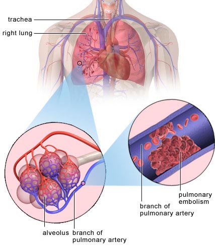
Example of where a pulmonary embolism may occur. Image: BUPA.


A pulmonary embolism is an obstruction or sudden blockage in the pulmonary artery or one of its branches, usually due to a blood clot that traveled to the lung from the leg. A clot that forms in one part of the body and travels in the bloodstream to another part of the body is called an embolus.


Pulmonary embolism (PE) is a serious condition that can cause:


  • Permanent damage to part of your lung from lack of blood flow to lung tissue
  • Low oxygen levels in your blood
  • Damage to other organs in your body from not getting enough oxygen
  •  

    If a clot is large, or if there are many clots, pulmonary embolism can cause death.


    In most cases, pulmonary embolism is a complication of a condition called deep vein thrombosis (DVT). In DVT, blood clots form in the deep veins of the body – most often in the legs. These clots can break free, travel to the lung, and block an artery.


    More than 600,000 people in the United States have a pulmonary embolism each year, and more than 60,000 of them die. Most of those who die do so within 30 to 60 minutes after symptoms start. Pulmonary embolism is one of the most common causes of death in hospitalized people who must remain in bed for a long time.


    Causes

    Nine out of 10 cases of pulmonary embolism are caused by blood clots that form in the legs (deep vein thrombosis) and then travel to the lungs.


    Leg clots can form when blood flow is restricted and slows down. This happens when you do not move around for long periods of time, such as:


  • After some types of surgeries
  • During a long trip in a car or on an airplane
  • If you must stay in bed for an extended time
  • Veins damaged from surgery or injured in other ways are more prone to blood clots.
  •  

    Rarely, an air bubble, part of a tumor, or other tissue travels to the lung and causes pulmonary embolism.


    Diagnosis

    Your doctor will take a health history and perform a physical exam in order to:


  • Identify your risk factors for deep vein thrombosis (DVT) and pulmonary embolism
  • See how likely it is that you could have a pulmonary embolism
  • Eliminate other possible causes for your symptoms
  •  


    Physical exam

    The physical exam will include:


  • Checking your blood pressure, heart rate, and breathing rate
  • Listening to your lungs
  • Listening to your heart
  • Checking your legs for signs and symptoms of DVT
  •  


    Tests

    To help determine whether you have a pulmonary embolism or related disease or condition, initial testing includes:


  • Electrocardiogram (EKG or ECG) to measure the rate and regularity of your heartbeat.

  • Chest X-ray, which takes a picture of the lungs, heart, large arteries, ribs, and the diaphragm.

  • Duplex ultrasound, the most common test used to diagnose deep vein clots. It uses sound waves to evaluate the flow of blood in your veins. A gel is put on the skin of the leg. A hand-held device is placed on the leg and passed back and forth over the affected area. This device sends sound waves from the leg to an ultrasound machine. A computer then turns the sounds into a picture. The picture is displayed on the computer screen, where your doctor can see the blood flow in your leg. This is an indirect way to diagnose a source of pulmonary embolism.

  • Blood work:
  •        To check for inherited disorders that cause clots.
           To measure the amount of oxygen and carbon dioxide in the blood (arterial blood gas). A pulmonary embolism may change the levels of oxygen and carbon dioxide in the blood.


    Your doctor will order additional tests based on how likely it is that you have a pulmonary embolism and which tests are available. Tests could include:


  • Ventilation-perfusion lung scan (VQ scan), a test that uses a radioactive material to see how well air and blood are flowing to all areas of the lung.

  • Pulmonary angiography, an accurate, invasive test used to diagnose pulmonary embolism. A flexible tube called a catheter is threaded through the upper thigh (groin) or arm to the blood vessels in the lung. Dye is injected through the catheter and then an X-ray picture is taken to show the blood flow through the blood vessels in the lungs. This test is not available at all hospitals, and a trained specialist is required to perform the test.

  • Spiral computed tomography (CT) scan, a special type of X-ray that takes pictures of structures inside the body. Dye is injected into a vein, allowing the doctors to see the blood vessels in the lungs on the X-ray picture. This is a very quick test and results are available shortly after the scan is completed.

  • Magnetic resonance imaging (MRI), a test that takes pictures of organs and structures inside the body. MRI uses radio waves to make the image. In many cases, an MRI can provide information that would not show on an X-ray.

  • Echocardiogram, a test that uses sound waves to check for blood clots inside the heart and to check heart function. It can also determine other possible causes for your symptoms.

  • D-dimer, a blood test that measures a substance in the blood that is released when a clot breaks up. High levels may indicate a clot. If your test is normal, a pulmonary embolism is not likely.
  •  


    Treatment

    The primary goals in treating pulmonary embolism are to:


  • Keep the blood clot or clots lodged in the lungs from getting bigger
  • Stop the development of new clots
  •  

    Treatment may include:


  • Medicines
  • Emergency treatment to dissolve a clot
  • Other treatments
  •  

    Treatment will vary depending on how severe your symptoms are. If your symptoms are severe, you need immediate emergency treatment. If your symptoms are mild, you can be treated as an outpatient.


    Medicines

    Anticoagulants (blood thinners) decrease your blood's ability to clot. They are used to stop clots from getting bigger and to prevent blood clots from forming. Anticoagulants do not break up blood clots that have already formed. Your body's natural system will dissolve the clot. Anticoagulants can be given:


  • As a pill (warfarin)
  • As an injection or shot (heparin)
  • In a vein (intravenous or IV) (heparin)
  •  

    Heparin and warfarin may be given at the same time. Heparin acts quickly, whereas warfarin takes 2 to 3 days before it starts to work. Once warfarin is working, the heparin will be stopped. Pregnant women cannot take warfarin and are treated with heparin only.


    If you have deep vein thrombosis, treatment with anticoagulants usually lasts for 3 to 6 months. However, the length of treatment may vary if:


  • Your blood clot occurred after a short-term risk, such as surgery. In this case, your treatment may be shorter.
  • You have had clots before. You will need longer treatment.
  • You are being treated for another illness (such as cancer). You will take anticoagulants for as long as those risk factors are present.
  •  

    The most common side effect of anticoagulants is bleeding. You need to have regular blood tests to check how well the medicine is working. You should call your doctor right away if you have easy bruising or bleeding.


    Thrombin inhibitors are new medicines that interfere with the clotting process. They are used to treat some types of clots for patients who cannot take heparin.


    Emergency treatment

    When pulmonary embolism is life threatening, doctors may use treatments to remove or break up clots in the lungs. These treatments are given in the emergency room or in the hospital and include:


  • Medicines called thrombolytics to quickly dissolve the blood clot. Thrombolytics are used to treat large clots causing severe symptoms.
  • Surgery to remove the blood clot.
  • A new procedure that uses a catheter to reach the clots. The catheter is inserted into the upper thigh (groin) or arm and threaded to the clot in the lung. The catheter may be used to extract the clot or deliver medicine to dissolve it.
  •  


    Additional considerations

    Vena cava filters are used when you cannot take medicines to thin your blood, or if you are taking blood thinners and continue to develop clots. The filter is inserted inside a large vein called the inferior vena cava (the vein that carries blood from the body back to the heart). It can catch the clots as they try to move through the body to the lungs. This treatment can prevent a clot from traveling to the lungs. It cannot stop other clots from forming.


    Graduated compression stockings are worn on the legs from the arch of the foot to just above or below the knee. These stockings are tight at the feet and become looser as they go up the leg. This causes a gentle compression (or pressure) up your leg. The stockings provide support and reduce the chronic swelling that can occur in the leg after a blood clot has occurred.


    Prevention

    Preventing pulmonary embolism begins with preventing deep vein thrombosis (DVT). It is important to know whether you are at risk for DVT and to take steps to lower your risk.


    If you have never had a deep vein clot, but are in a situation that may increase your risk, be sure to:


  • Exercise your lower leg muscles during long car trips and plane rides.
  • Get out of bed and move around as soon as you are able after having surgery or being ill. The sooner you move around, the lower the chance you have of developing a clot.
  • Take medicines to prevent clots after some types of surgery as directed by your doctor. Follow up with your doctor.
  •  

    If you already have had DVT or a pulmonary embolism, you need to prevent further clots from developing by:


  • Seeing your doctor for regular checkups
  • Taking your medicines to prevent or treat blood clots as prescribed by your doctor
  • Using compression stockings as directed to prevent chronic swelling in your legs after a DVT, if prescribed by your doctor
  • Contacting your doctor immediately if you have any signs or symptoms of DVT or pulmonary embolism
  •  


    Life after pulmonary embolism

    Once you have had a pulmonary embolism (with or without deep vein thrombosis (DVT)), you have a greater chance of having another one. After treatment has begun and you are feeling better, your treatment will continue at home.


    At home, you will continue taking blood-thinning medicines. It is important that you:


  • Take medicines as prescribed.
  • Check with your doctor before taking any other medicines, including over-the-counter medicines, vitamins, or herbal treatments.
  • Continue to have regular blood tests as directed by your doctor.
  • Check with your doctor before changing your diet or taking any supplements.
  • Wear your compression stockings as directed, if prescribed by your doctor.
  •  

    Medicines used to treat pulmonary embolism can thin your blood too much. This can cause:


  • Bleeding in the digestive system. Get immediate treatment if you:
  •       Vomit bright red blood or material that looks like coffee grounds
          Pass bright red blood in your stool or have black tarry stools
          Develop pain in your abdomen


  • Excessive bleeding from a fall or injury. Get immediate treatment if you cannot stop any bleeding after applying pressure for 10 minutes.
  •  

  • Bleeding in the brain. Get immediate treatment if you:
  •       Develop a severe headache
          Have any sudden changes in your vision
          Have sudden loss of movement of your legs or arms
          Have sudden memory loss or confusion


    Treatment for pulmonary embolism usually lasts up to 6 months. During treatment and after, you should continue to:


  • Prevent DVT
  • Check your legs for any signs or symptoms of DVT