
ankle
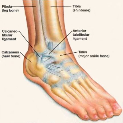
The ankle joint is the hinge joint between the foot and the leg. The uppermost bone of the foot, called the talus (ankle-bone), fits between the two bony protuberances formed by the lower ends of the tibia (shin bone) and fibula.
The ankle joint has great stability. The deep mortise formed by the lower end of the tibia and the medial and lateral malleoli securely holds the talus in position. Strong ligaments on each side of the ankle joint provide support and limit movement. On the outside of the ankle are three main ligaments called the anterior talofibular ligament (connecting the talus and the fibula at the front), the calcaneofibular ligament (connecting the calcaneus bone to the talus), and the posterior talofibular ligament (connecting the talus and fibula at the back). Holding the tibia to the talus bone on the inside of the ankle is the broad, fan shaped deltoid ligament.
The ankle joint allows up-and-down movements of the foot. Other movements of the foot, such as tilting and rotating, occur at joints in the foot itself.
Ligaments of the ankle
The medial, or deltoid, ligament is very strong and is attached by its apex to the tip of the medial malleolus. Below, the deep fibers are attached to the nonarticular area on the medial surface of the body of the talus; the superficial fibers are attached to the medial side of the talus, the sustentaculum tali, the plantar calcaneonavicular ligament, and the tuberosity of the navicular bone.
The lateral ligament is weaker than the medial ligament and consists of three distinct bands.
Movements of the ankle
The movements of the ankle are dorsiflexion (toes pointing upward) and plantar flexion (toes pointing downward). The movements of inversion and eversion take place at the tarsal joints and not at the ankle joint.
Dorsiflexion is performed by the tibialis anterior, extensor hallucis longus, extensor digitorum longus, and peroneus tertius. It is limited by the tension of the tendo calcaneus, the posterior fibers of the medial ligament, and the calcaneofibular ligament.
Plantar flexion is performed by the gastrocnemius, soleus, plantaris, peroneus longus, peroneus brevis, tibialis posterior, flexor digitorum longus, and flexor hallucis longus. It is limited by the tension of the opposing muscles, the anterior fibers of the medial ligament, and the anterior talofibular ligament.
Note that during dorsiflexion of the ankle joint, the wider anterior part of the articular surface of the talus is forced between the medial and lateral malleoli, causing them to separate slightly and tighten the ligaments of the inferior tibiofibular joint. This arrangement greatly increases the stability of the ankle joint when the foot is in the initial position for major thrusting movements in walking, running, and jumping.
Note also that when the ankle joint is fully plantar flexed, the ligaments of the inferior tibiofibular joint are less taut and small amounts of rotation, abduction, and adduction are possible.
Sprains and fracture dislocations of the ankle
Sprains of the ankle are usually caused by excessive inversion of the foot. The anterior talofibular ligament and the calcabeofibular ligament are partially torn, causing great pain and local swelling. A similar but less common injury may occur to the medial or deltoid ligament as the result of excessive eversion. The great strength of the medial ligament usually results in the ligament pulling off the tip of the medial malleolus.
Fracture dislocations of the ankle joint are common. Forced external rotation and over-eversion of the foot is the most common cause of fracture dislocation. The talus is externally rotated forcibly against the lateral malleolus of the fibula. The torsion effect on the lateral malleolus causes it to fracture spirally. If the force continues, the talus moves laterally, and the medial ligament of the ankle joint becomes taut and pulls off the tip of the medial malleolus. If the talus is forced to move still farther, its rotary movement results in its violent contact with the posterior inferior margin of the tibia, which shears off.
Other less common types of fracture dislocation are due to forced over-eversion (without rotation), in which the talus presses the lateral malleolus laterally and causes it to fracture transversely. Over-inversion (without rotation), in which the talus presses against the medial; malleolus, will produce a vertical fracture through the base of the medial malleolus.

