
coronary calcium scan
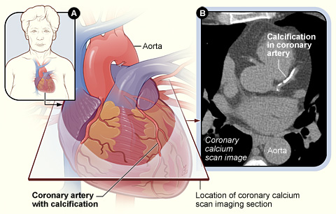
Figure A shows the position of the heart in the body and the location and angle of the coronary calcium scan image. Figure B is the coronary calcium scan image showing calcification in a coronary artery.
A coronary calcium scan is a test that can help show whether you have coronary artery disease (CAD). In CAD, a fatty material called plaque narrows your coronary (heart) arteries and limits blood flow to your heart. CAD is the most common type of heart disease in both men and women. It can lead to angina, heart attack, heart failure, and arrhythmia.
Coronary calcium scanning looks for specks of calcium (called calcifications) in the walls of the coronary arteries. Calcifications are an early sign of heart disease. The test can show, before other signs and symptoms occur, whether you're at increased risk for a heart attack or other heart problems.
A coronary calcium scan is most useful for people who are at moderate risk for a heart attack. You or your doctor can calculate your 10-year risk using the Risk Assessment Tool from the National Cholesterol Education Program. People at moderate risk have a 10 to 20 percent chance of having a heart attack within the next 10 years. The coronary calcium scan helps doctors decide who within this group needs treatment.
Two machines can show calcium in the coronary arteries – electron beam computed tomography (EBCT) and multidetector computed tomography (MDCT). Both use an X-ray machine to make detailed pictures of your heart. Doctors study the pictures to see whether you're at risk for heart problems in the next 2 to 10 years.
A coronary calcium scan is simple and easy for the patient, who lies quietly in the scanner machine for about 10 min. Pictures of the heart are taken that show whether the coronary arteries have calcifications.
Before a cardiac calcium scan
No special preparation is needed. You may be asked to avoid caffeine and smoking for 4 hours before the test. For the scan, you will remove your clothes above the waist and wear a hospital gown. You also will remove any jewelry from around your neck or chest.
During cardiac calcium scan
Coronary calcium scans are done in a hospital or outpatient office. The X-ray machine that's used is called a computed tomography (CT) scanner.
The technician who operates the scanner will clean areas of your chest and apply small sticky patches called electrodes. The electrodes are attached to an EKG (electrocardiogram) monitor. The EKG measures the electrical activity of your heart during the scan. This makes it possible to take pictures of your heart when it's relaxed, between beats.
The CT scanner is a large machine that has a hollow, circular tube in the center. You will lie on your back on a sliding table. The table can move up and down and goes inside the tunnel-like machine.
The table will slowly slide into the opening in the machine. Inside the scanner, an X-ray tube moves around your body to take pictures of your heart. You may be asked to hold your breath for 10 to 20 sec while the pictures are taken. This prevents movement in the image.
During the test, the technician will be in a nearby room with the computer that controls the CT scanner. The technician can see you through a window and talk to you through an intercom system.
You may be given medicine to slow down a fast heart rate. This helps the machine take better pictures of your heart. The medicine will be given by mouth or injected into a vein.
A coronary calcium scan takes about 5 to 10 min. During the test, the machine makes clicking and whirring sounds as it takes pictures. It causes no discomfort, but the exam room may be chilly to keep the machine working properly.
If you become nervous in enclosed spaces, you may need to take medicine to stay calm. This isn't a problem for most people, because your head will remain outside the opening in the machine.
After cardiac calcium scan
You're able to return to your normal activities after the coronary calcium scan is done. A doctor who is trained in reading these scans will discuss the results with you.
What a cardiac calcium scan shows
After the coronary calcium scan, you will get a calcium score called an Agatston score. The score is based on the amount of calcium found in your coronary arteries. You may get an Agatston score for each major artery and a total score.
The test is negative if no sign of calcium deposits (calcifications) is found in your arteries. This means your chance of having a heart attack in the next 2 to 5 years is low.
The test is positive if calcifications are found in your arteries. Calcifications are a sign of atherosclerosis and coronary artery disease. (Atherosclerosis is when the arteries harden and narrow due to plaque buildup.) The higher your Agatston score, the greater the amount of atherosclerosis.
Risks
Coronary calcium scanning has very few risks. The test isn't invasive, which means that no surgery is done and no instruments are inserted into your body. Coronary calcium scanning doesn't require an injection of contrast dye to make your heart or arteries visible on the X-ray images.
Because an X-ray machine is involved, you will be exposed to a small amount of radiation. Electron-beam computed tomography (EBCT) uses less radiation than multidetector computed tomography (MDCT). In either case, the amount of radiation is less than or equal to the amount of radiation you're naturally exposed to in a single year.

