
organ of Corti
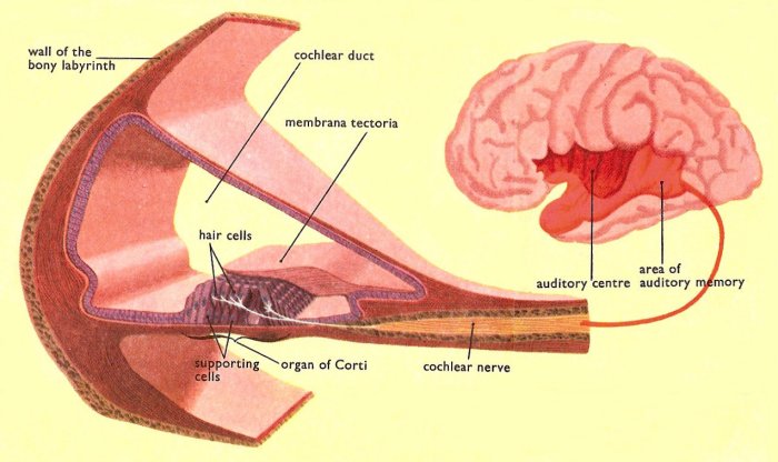
Sectional view showing how the parts of the organ of Corti are arranged within the cochlear duct.
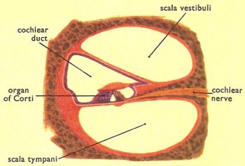
Cross-section of one spiral of the cochlea
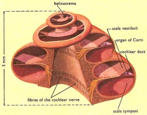
Cutaway illustration of the cochlea.
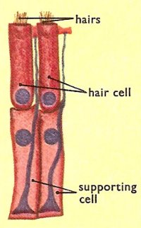
Two hair cells and two supporting cells shown as they are arranged in the organ of Corti.
The organ of Corti lies within the cochlea of the inner ear. In the organ of Corti, sound vibrations which pass along the cochlear duct are converted into nerve impulses. These impulses are transmitted along the cochlear nerve, or auditory nerve, to the brain, where they are interpreted as sound.
The organ of Corti lies on the basilar membrane close to the attachment to the osseus lamina. In this situation it extends throughout the whole of the two and three-quarter turns of the cochlear duct.
Basically, the organ of Corti consists of two rows of rod cells which are arranged on the membrane to form a minute arch. To this arch are fixed four rows of hair cells, one row on the inner side and three on the outer. On both sides of the arch the hair cells are held firmly in position by rows of supporting cells. The membrana tectoria arches over the whole of the organ.
At the upper and free end of each hair a small number of acoustic hairs project into the cochlear duct. To each cell a minute fiber of the cochlear or acoustic nerve is attached.

