
neuron
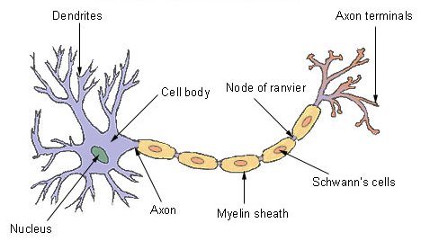
Figure 1. General structure of a typical neuron.
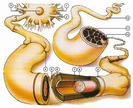
Figure 2. The structure of a single nerve and myelinated cell fiber is shown. (1) Cell body. (2) Dendrites conducting messages to the cell body. (3) Cell nucleus. (4) Node of Ranvier, a constriction of the myelin sheath. (5) Schwann cell nucleus – the cell secreting the myelin sheath (6) that insulates the axon (7). The latter conducts impulses from the cell body. (8) Nerve fiber. (9) Endoneurium – inter-fiber tissue. (10) Perineurium, sheathing fiber bundles. (11) Epineurium sheathing the nerve (12).
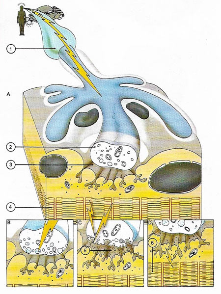
Figure 3. Nerve impulses are transmitted by bioelectricity. At the junction between nerve and muscle fibers (the motor end plate) a chemical transmitter is released from the nerve in response to the electrical change produced when the nerve is stimulated. As a result, the muscle fiber contracts. (A) shows a longitudinal section through a motor end plate. (1) shows a nerve axon, with an impulse travelling towards its tip. (2) shoes a section through a terminal button of a nerve cell, the part containing acetylcholine, a chemical transmitter. (3) is the synaptic gutter – a muscle fiber membrane greatly folded to form a 'gutter' that accommodates the terminal button. (4) is the muscle fiber itself in a relaxed state. (B) is a detailed diagram of the terminal button region, showing the nerve cell about to release the chemical transmitter. (C) demonstrates the release of the chemical transmitter (5) as the nerve impulse reaches the tip of the terminal button. The transmitter causes changes in the permeability of the muscle fiber membrane. (D) shows the contraction of the muscle fiber. The permeability change results in an altered electrical state across the membrane stimulating muscle fiber contraction. The muscle fiber filaments pull the fiber units closer together.
A neuron, or nerve cell, is a cell that supports the functions of the nervous system by reacting to stimuli and transmitting nerve impulses. It is highly specialized and amitotic. The term "amitotic" means that if a neuron is destroyed it cannot be replaced, because neurons do not undergo mitosis.
Each neuron consists of three basic parts: a cell body (soma) that contains the nucleus; one or more dendrites, which are short branches off the body that receive incoming impulses; and a single, long axon that carries impulses away from the body and to the next neuron (Figures 1 and 2).
Cell body
In many ways, the cell body is similar to other types of cells. It has a nucleus with at least one nucleolus and contains many of the typical cytoplasmic organelles. It lacks centrioles, however. Because centrioles function in cell division, the fact that neurons lack these organelles is consistent with the amitotic nature of the cell.
Dendrites
Dendrites and axons are cytoplasmic extensions, or processes, which project from the cell body. They are sometimes referred to as fibers. Dendrites are usually, but not always, short and branching, which increases their surface area to receive signals from other neurons. The number of dendrites on a neuron varies. They are called afferent processes because they transmit impulses to the neuron cell body. There is only one axon that projects from each cell body. It is usually elongated and because it carries impulses away from the cell body, it is called an efferent process.
Axon
main article
An axon may have infrequent branches called axon collaterals. Axons and axon collaterals terminate in many short branches or telodendria. The distal ends of the telodendria are slightly enlarged to form synaptic bulbs. Many axons are surrounded by a segmented, white, fatty substance called myelin or the myelin sheath. Myelinated fibers make up the white matter in the central nervous system (CNS), while cell bodies and unmyelinated fibers make the gray matter. The unmyelinated regions between the myelin segments are called the nodes of Ranvier.
In the peripheral nervous system, the myelin is produced by Schwann cells. The cytoplasm, nucleus, and outer cell membrane of the Schwann cell form a tight covering around the myelin and around the axon itself at the nodes of Ranvier. This covering is the neurilemma, which plays an important role in the regeneration of nerve fibers. In the central nervous system (CNS), oligodendrocytes produce myelin, but there is no neurilemma, which is why fibers within the CNS do not regenerate.
Functionally, neurons are classified as afferent, efferent, or interneurons (association neurons) according to the direction in which they transmit impulses relative to the central nervous system. Afferent, or sensory, neurons carry impulses from peripheral sense receptors to the CNS (see sensory nerve). They usually have long dendrites and relatively short axons. Efferent, or motor, neurons transmit impulses from the CNS to effector organs such as muscles and glands (see motor neuron). Efferent neurons usually have short dendrites and long axons. Interneurons, or association neurons, are located entirely within the CNS in which they form the connecting link between the afferent and efferent neurons. They have short dendrites and may have either a short or long axon.
Glial cells
Although the nervous system is very complex, there are only two main types of cells in nerve tissue. The actual nerve cell is the neuron. It is the "conducting" cell that transmits impulses and the structural unit of the nervous system. The other type of cell is the glial cell, or neuroglia. The word "neuroglia" means "nerve glue." Neuroglia cells do not conduct nerve impulses, but instead, they support, nourish, and protect the neurons. They are a special type of connective tissue for the nervous system. Glial cells are far more numerous than neurons and, unlike neurons, are capable of mitosis.
Glial cells not only provide physical support but also respond to injury, regulate the chemical environment of neurons, form the myelin insulation of nervous pathways, help guide neuronal migration during development, and play an important role in the blood-brain barrier. Glial cells come in four main types:
· astrocytes
· oligodendrocytes
· ependymal cells
· microglia
Nerves
Nerves consist of bundles of neurons. More specifically, a nerve is a bundle of axons, with accompanying connective tissue and blood vessels. Nerves use chemical and electrical signals to transmit sensory and motor information from one part of the body to another. Each nerve fiber conducts impulses independently of its neighbors. See also cranial nerve and spinal nerve.
Nerve impulse
A nerve impulse (Figure 3) is an electrical signal that travels along the axon of a neuron. Nerve impulses carry information throughout the nervous system. As an impulse passes, sodium and potassium ions flow into and out of the axon's membrane (see sodium-potassium pump), creating a voltage reduction called the action potential. Nerve impulses can travel at up to 150 meters per second (500 feet per second).
Action potential
Action potential is an all-or-nothing change in electrical potential that occurs across the plasma membrane of a nerve cell during the passage of a nerve impulse. As an impulse travels along the axon of a nerve cell, it causes a localized and transient switch in potential across the membrane from about –60 millivolts (the so-called resting potential) to +45 millivolts. The change in potential is caused by an influx of sodium ions. Nervous stimulation of muscle cells and some glandular cells has a similar effect.
Resting potential
Resting potential is the potential difference between the two sides of the membrane of a nerve cell when the cell is not conducting a nerve impulse.

