
thyroid cartilage
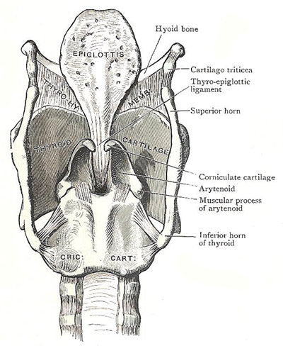
Posterior aspect of cartilages and ligaments of the larynx.
The thyroid cartilage is of the hyaline variety and may show early ossification. It is the largest of the laryngeal cartilages; and the relatively larger size of the larynx in men – which is a well known secondary sex character – is manifested best by the thyroid cartilage. It is in the form of a pair of broad, quadrilateral laminae which are fused together in front but are widely separated behind.
The anterior borders of the laminae are fused only in their lower parts. Above, they are separated by a deep, narrow V-shaped thyroid notch. In men, the angle formed by the meeting of the anterior borders of the two laminae, especially in the upper part, is very projecting; and, with the margins of the notch, it makes the marked, subcutaneous laryngeal prominence, colloquially known as Adam's apple.
The posterior border of each lamina is thick and rounded, and is prolonged upwards and downwards as two processes termed the horns. The superior horn gives attachment to the lateral thyrohyoid ligament. The inferior horn is shorter and thicker and curves slightly medially, and the medial side of its tip articulates with the side of the cricoid cartilage.
The superior border of the lamina is for the most part slightly convex, and anteriorly it dips down to become continuous with the margin of the thyroid notch. The inferior border is almost horizontal, and has a small projection on it, the inferior tubercle, a little behind its middle.
The lateral surface of the lamina is relatively flat. Immediately below the posterior part of the upper border, and in front of the root of the superior horn, there is a prominence called the superior tubercle. From that point an oblique ridge decends towards the inferior tubercle. The ridge gives attachment to the sternothyroid and thyrohyroid muscles and to the inferior constrictor of the pharynx, which arises also from the area behind the ridge.
The medial surface of the lamina is smooth and slightly concave. It is lined with mucous membrane, and several structures are attached to it at and near the anterior border – the thyroepiglottic ligament, and vestibular and vocal ligaments, and the thyroarytenoid and vocalis muscles.

