
cardiac MRI
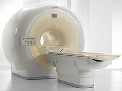
Cardiac MRI unit. Credit: Philips.
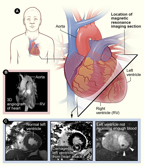
Figure A shows the heart's position in the body and the location and angle of the MRI images shown in figure C. Figure B is a MRI angiogram, which is sometimes used instead of a standard angiogram. Figure C shows MRI pictures of a normal left ventricle (left image), a left ventricle damaged from a heart attack (middle image), and a left ventricle that isn't getting enough blood from the coronary arteries (right image).
Magnetic resonance imaging (MRI) is a safe, noninvasive test that creates detailed images of your organs and tissues. "Noninvasive" means that no surgery is done and no instruments are inserted into your body.
MRI uses radio waves and magnets to create images of your organs and tissues. Unlike computed tomography scans (also called CT scans) or conventional X-rays, MRI imaging doesn't use ionizing radiation or carry any risk of causing cancer.
Cardiac MRI uses a computer to create images of your heart as it's beating, producing both still and moving pictures of your heart and major blood vessels. Doctors use cardiac MRI to get images of the beating heart and to look at the structure and function of the heart. These images can help them decide how best to treat patients with heart problems.
Cardiac MRI is a common test for diagnosing and evaluating a number of diseases and conditions, including:
Cardiac MRI images can help explain results from other tests, such as X-ray and CT scans. Cardiac MRI is sometimes used to avoid the need for other tests that use radiation (such as X-rays), invasive procedures, and dyes containing iodine (these dyes may be harmful to people who have kidney problems).
Sometimes during cardiac MRI, a special dye is injected into a vein to help highlight the heart or blood vessels on the images. Unlike the case with X-rays, the special dyes used for MRI don't contain iodine, so they don't present a risk to people who are allergic to iodine or have kidney problems.
Before cardiac MRI
You'll be asked to fill out a screening form before the test takes place. The form may ask whether you've had previous surgeries, whether you have any metal objects in your body, and whether you have any medical devices (like a cardiac pacemaker) surgically implanted in your body.
Most, but not all, implanted medical devices are allowed near the magnetic resonance imaging (MRI) machine. Ask your doctor or the technician operating the machine if you have concerns about any implanted devices or conditions that may interfere with the MRI.
MRI can interfere seriously with some types of implanted medical devices.
Your doctor will let you know if you shouldn't have a cardiac MRI because of a medical device.
Your doctor or technician will tell you whether you need to change into a hospital gown for the test. Don't bring hearing aids, credit cards, jewelry and watches, eyeglasses, pens, removable dental work, and anything that's magnetic near the MRI machine.
Tell your doctor if you have a history of becoming anxious or fearful when in a fairly tight or confined space. This fear is called claustrophobia. In this case, your doctor might give you medicine to help you relax. Your doctor may ask you to stop eating 6 hours before you take this medicine on the day of the test.
Some of the newer cardiac MRI machines are open on all sides. Ask your doctor to help you find a facility that has an open MRI machine if you're fearful in tight or confined spaces.
Your doctor will let you know if you need to arrange for a ride home after the test.
During cardiac MRI
Magnetic resonance imaging (MRI) machines are usually located at a hospital or at a special medical imaging facility. A radiologist or other physician with special training in medical imaging oversees MRI testing.
Cardiac MRI usually takes 45 to 90 minutes, depending on how many images are needed. The test may take less time with some newer MRI machines.
The MRI machine will be located in a specially constructed room. This will prevent radio waves from disrupting the machine and will prevent the strong magnetic fields generated by the MRI machine from interfering with other equipment.
Traditional MRI machines look like a long, narrow tunnel. Newer MRI machines called short-bore systems are shorter, wider, and don't completely surround you. Some of the newer machines are open on all sides. Your doctor will help decide which machine type is best for you.
Cardiac MRI is painless and harmless. You will lie on your back on a sliding table that goes inside the tunnel-like machine. The technician will control the machine from the next room. He or she will be able to see you through a glass window and will be able to talk to you through an intercom system. Tell the technician if you have a hearing problem.
The MRI machine makes loud humming, tapping, and buzzing noises. Earplugs may help lessen the noises made by the MRI machine. Some facilities let you listen to music during the test.
Remaining very still during the test is important. Any movement may blur the images. If you're unable to lie still, you may be given medicine to help you relax. You may be asked to hold your breath for 10 to 15 seconds at a time while the technician takes pictures of your heart. Researchers are studying ways that will allow someone having a cardiac MRI to breathe freely during the exam, while achieving the same image quality.
A contrast dye, such as gadolinium, may be used to highlight your blood vessels or heart in the images. Contrast dye is usually injected into a vein in your arm with a needle. You may feel a cool sensation during the injection, and you may feel discomfort where the needle was inserted. Gadolinium doesn't contain iodine so it won't create problems for people who are allergic to iodine.
If your cardiac MRI includes a stress test to detect blockages in your coronary arteries, you will receive other medicines to increase the blood flow in your heart or to increase how fast your heart beats.
After cardiac MRI
Once the cardiac magnetic resonance imaging (MRI) is done, if you haven't received medicine to help you relax, you will be able to return to normal activities immediately.
If you did receive medicine to help you relax during the test, your doctor will tell you when you can return to normal activities. You will need someone to drive you home.
What does cardiac MRI show?
The doctor supervising your scan will provide your doctor with the results of your cardiac MRI. Your doctor will discuss the findings with you.
Cardiac MRI can reveal various heart conditions and disorders, such as:
Cardiac MRI is a fast, accurate tool that can help diagnose a heart attack by detecting areas of the heart that don't move normally, have poor blood supply, or are scarred. Cardiac MRI can show whether any of the coronary arteries are blocked, causing reduced blood flow to your heart muscle.
Currently, coronary angiography is the procedure most commonly used to look at blockages in the coronary arteries. Coronary angiography is an invasive procedure that uses X-rays and iodine-based contrast dye. Researchers have found that cardiac MRI can replace coronary angiography in some cases, avoiding the need to use X-ray radiation and iodine-based dyes.
Researchers are discovering new ways to use cardiac MRI. In the future, cardiac MRI may be able to replace X-rays as the main way to guide invasive procedures such as cardiac catheterization. Also, improvements in cardiac MRI are likely to lead to better methods for detecting heart disease in the future.
Risks
Cardiac magnetic resonance imaging (MRI) produces no side effects from the magnetic fields and radio waves. This method of obtaining images of organs and tissues doesn't carry a risk of causing cancer or birth defects.
Serious reactions to the special contrast dyes used for MRI are very rare. However, side effects are possible and include the following:
Rarely, the contrast dyes can be harmful in patients with severe kidney disease.
If your cardiac MRI includes a stress test, more medicines will be used during the test. These medicines may have additional side effects that aren't expected during a regular MRI scan, such as:

