
cavernous sinus
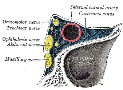
Oblique section through the cavernous sinus.
The cavernous sinus is an important sinus, not as a venous channel (although its connection with the ophthalmic veins gives it a measure of importance), but because of its intimate relationship to important parts – to the sphenoidal air sinus, the hypophis, the internal carotid artery the oculomotor, trochlear, and abducent nerves, the ophthalmic and maxillary divisions of the trigeminal nerve, and the trigeminal ganglion.
The cavernous sinus is a short, wide channel that lies on the side of the body of the sphenoid bone, extending from the medial end of the superior orbital fissure to the apex of the petrous temporal bone. Like the other venous sinuses, it is formed by the separation of the two layers of the dura mater and is lined with endothelium; but its cavity is traversed by numbers of delicate, interlacing fibrous threads that break it up into a large number of intercommunicating "caverns", from which the sinus derives its name.
Tributaries and communications
At its anterior end the cavernous sinus receives the ophthalmic vein or veins and the sphenoparietal sinus, which lies along the edge of the lesser wing of the sphenoid. Its posterior end is connected with the superior and inferior petrosal sinuses. Medially, a number of small intercavernous sinuses connect it with its fellow; one of them passes through the anterior margin of the diaphragma sellae, another through the posterior margin, and the others pass below the hypophis. Superiorly, it receives one or more small cerebral veins and one large one – the superficial middle cerebral vein. Inferiorly, it communicates with venous plexuses outside the skull by means of emissary veins: one emissary vein passes through the carotid canal to connect it with the pharyngeal plexus; another passes through the foramen ovale or through the emissary sphenoidal foramen, when that foramen is present, and connects it with the pterygoid plexus.
Relations
The relations of the cavernous sinus are important. The sphenoidal air sinus is below and medial to it. The hypophis is closely related to its upper margin. The trigeminal ganglion is immediately lateral to its posterior end. The maxillary nerve runs forward along its lower border. The oculomotor, trochlear, and ophthalmic nerves are embedded in its lateral wall. The internal carotid artery and the abducent nerve are in the lower part of the sinus, shut off from its cavity merely by the endothelium.

