lens (eye)

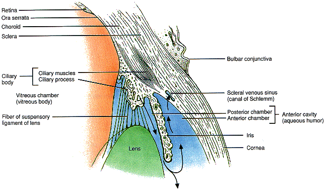
Anatomy of the lens and surrounding structures.
The lens is a transparent structure located just behind the pupil of the vertebrate eye. It lies in the aqueous humor, by which it is oxygenated and nourished, and is attached by suspensory ligaments to the ciliary body. The lens consists largely of elongated inert cells (lens fibers), which contain large amounts of special lens proteins called crystallins.
In land-dwelling animals, including man, the lens of the eye is not as important as the cornea in the process of refraction (bending of light) which produces images on the retina. Rather its main function is accommodation by change of shape. In fish, by contrast, the lens of the eye is spherical and is responsible for both refraction and accommodation.
In the embryonic development of vertebrates, the lens is derived from the epidermis, as an induction by the retina.
Lens dislocation
Displacement of the lens from its normal position is almost always caused by an injury that ruptures some or all of the fibers that connect the lens to the ciliary body. In Marfan syndrome, the fibers are particularly weak and lens dislocation is common.
A dislocated lens may slide sideways, upwards, or downwards, causing severe visual distortion or double vision in the affected eye, or it may slip backwards into the vitreous humor. A lens dislocated forwards through the pupil usually causes a form of glaucoma because of closure of the drainage angle of the eye.
Lenticonus
A condition on which the central part of the front surface of the lens of the eye (or sometimes, the back) has a much steeper curvature than normal and bulges forwards in a blunted cone. It is usually congenital.
Surgical removal of the lens is called lensectomy.


