
retina
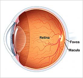
Figure 1. Inside the human eye. Credit: Mayo Clinic.
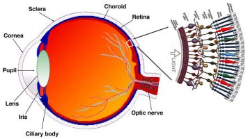
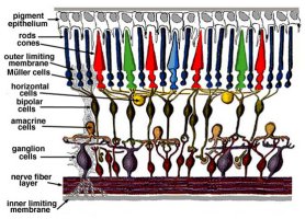
Figure 2. The layers that make up the retina. Credit: John Moran Eye Center, University of Utah.
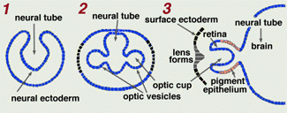
Figure 3. Development of the eye.
The retina is the light-sensitive layer that lines the interior of the eye of vertebrates and some cephalopods. In the vertebrate eye it is part of the central nervous system – effectively a photosensitive extension of the brain.
Anatomy of the human retina
The retina is roughly 0.5 mm thick and forms about three-quarters of a full sphere (being open at the front where light enters the eye). In the center of the retina is the optic disk, a circular to oval white area measuring about 2 × 1.5 milimeters across. This disk is where the axons of ganglion cells leave the eye to form the optic nerve. Because there are no light-sensitive cells in the optic disk, this part of the eye is also known as the blind spot. From the center of the optic disk radiate the major blood vessels of the retina. Displaced temporally (in the direction of the temples) from the optic disk is the macula, in the middle of which is the fovea, responsible for detailed vision (Figure 2).
The central part of the retina is dominated by cone cells, whereas the peripheral retina is dominated by rod cells. Altogether there are about 7 million cones and 100 million rods.
Like all vertebrate retinas, the human retina has three layers of nerve cell bodies and two layers of synapses. Through a quirk of vertebrate evolution, the ganglion cells lie innermost in the retina while the light-sensitive rods and cones are outermost, so that light first has to pass through the thickness of the retina before it reaches the photoreceptive cells.
Embryology of the retina
Embryologically the retina is part of the brain. It develops as a hollow outpushing of the neural tube, whose outer end becomes indented to form a stalked cup; the double-walled cup forms the two layers of the retina (Figure 3).
In cephalopods the retina is formed not as an outgrowth of the brain but from the external ectoderm and the light-sensitive cells are external to the nerve fibers. This reveals that, although vertebrate and cephalopod eyes have some striking similarities, they are biologically analogous rather than homologous.
Comparison of vertebrate and cephalopod retinas
As mentioned, the vertebrate retina is back-to-front, or inverted, in that the light-sensing cells are on the back side of the retina, so that light has to travel through a layer of neurons before it gets to the photoreceptors. By contrast, the cephalopod retina is everted: the photoreceptors lie at the front of the retina, with processing neurons behind them. Because of this arrangement, cephalopods do not have a blind spot.

