
mandibular nerve
The mandibular nerve is the largest of the three divisions of the trigeminal nerve. It arises, within the cranium, from the lower part of the convex border of the trigeminal ganglion, and enters the infratemporal fossa through the foramen ovale. Until it leaves the skull it is composed (like the ophthalmic and maxillary branches of the trigeminal) wholly of sensory fibers. But, as it descends through the foramen ovals, it is joined by the motor root of the trigeminal nerve; and, below the skull, it is therefore a mixed nerve.
Immediately after its exit from the skull, the mandibular nerve gives off the nervus spinosus and the nerve to the medial pterygoid muscle, and, at a slightly lower level, it divides into an anterior division and a posterior division.
The undivided trunk lies immediately below the skull, on the medial side of the lateral pterygoid, behind the medial pterygoid, on the lateral side of the pharynx, where the tensor palati separates it from the pharyngotympanic tube; the middle meningeal artery is a little behind it.
The anterior division is composed chiefly of motor fibers. It divides almost at once into the deep temporal nerves, the nerves to the masseter and lateral pterygoid, and the buccal nerve; all its sensory fibers pass into the buccal nerve.
The posterior division gives off the two roots of the auriculotemporal nerve and then divides into the lingual and the inferior dental. The only motor fibers in it are those that form the mylohyoid branch of the inferior dental.
The nervus spinosus is a very slender branch which enters the cranium with the middle meningeal artery through the foramen spinosum. It supplies the dura mater, and sends a filament into a middle ear.
The nerve to the medial pterygoid passes forwards to enter the deep surface of the muscle. The otic ganglion is in close relation to its commencement.
The buccal nerve is the largest branch of the anterior division. It passes between the two heads of the lateral pterygoid muscle, and then downwards and forwards over the lower head to reach the surface of the buccinator muscle. After dividing and forming a plexus with the buccal branches of the facial nerve (which supplies the motor fibers to the buccinator), it supplies the skin and the mucous membrane of the soft part of the cheek. In its course downwards, it is opposite the anterior margin of the ramus of the mandible in close relation to the deep surface of the temporal muscle – parallel with its tendinous fasciculi and within its sheath. It is often in a groove flush with the deep surface of the muscle, and may even be within its substance.
The nerve to the lateral pterygoid arises in common with the buccal nerve and leaves it to enter the muscle as it passes between its heads. The deep temporal nerves – an anterior and a posterior – pass laterally above the lateral pterygoid and then upwards into the temporal fossa to sink into the temporal muscle; in their course to the muscle they closely to the bone. A deep temporal branch often arises from the buccal nerve after it has appeared between the heads of the lateral pterygoid. The branch passes upwards over the upper head of the lateral pterygoid to reach the temporal muscle; and it may replace the anterior deep temporal.
The nerve to the masseter arises in common with the posterior deep temporal. It passes laterally above the lateral pterygoid muscle and through the mandibular notch close behind the temporal muscle, and sinks into the upper part of the deep surface of the masseter near its posterior border. Before it reaches the masseter, it gives one or two twigs to the capsule of the mandibular joint.
The auriculotemporal nerve springs by two roots from the posterior division of the mandibular nerve, under cover of the lateral pterygoid. The two roots are composed of sensory fibers and each receives a communication from the otic ganglion, by means of which it is brought, indirectly, into association with the glossopharyngeal nerve. The roots embrace the middle meningeal artery, and unite behind it to form a stem which runs backwards deep to the neck of the mandible. At the interval between the ear and the mandible it turns upwards in close relation to the parotid gland, crosses the posterior root of the zygoma with the superficial temporal artery, and enters the temple, where it breaks up into terminal branches.
Its branches are: (1) A few slender filaments to the posterior part of the capsule of the mandibular joint. (2) One or two thick branches which enter the parotid gland to supply the gland and to join the upper branches of the facial nerve. (3) Auricular branches. (4) Terminal filaments to the skin over the temporal region.
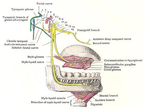
The inferior dental nerve is the largest branch of the posterior division of the mandibular nerve. It emerges from under cover of the lateral pterygoid at the lower border of the muscle, passes downwards on lateral surface of the sphenomandibular ligament and medial pterygoid muscle, and enters the mandibular foramen. The inferior dental branch of the maxillary artery runs downwards behind it, and the lingual nerve is in front of it, on a rather deeper plane. It runs downwards and forwards in the canal in the body of the mandible, gives off branches to the teeth of the lower jaw, and sends the mental nerve out through the mental foramen to supply the skin of the chin and the skin and mucous membrane of the lower lip.
The inferior dental nerve is a sensory nerve, but a few motor fibers are contained within its sheath, and separate off as the mylohyoid nerve.
The mylohyoid nerve arises near the mandibular foramen, pierces the sphenomandibular ligament and proceeds downwards and forwards, in a groove on the medial surface of the mandible, to the digastric triangle. In the digastric triangle, where it is joined by the submental artery, the nerve breaks up into numerous branches for the supply of the mylohyoid muscle, and the anterior belly of the digastric.
The lingual nerve is almost as thick as the inferior dental. It is entirely sensory and is distributed to the mucous membrane of the anterior two-thirds as the tongue. In the first part of its course, like the other branches of the mandibular nerve, it is medial to the lateral pterygoid muscle. As it descends it appears at the lower border of the muscle in front of the inferior dental nerve. It then proceeds downwards and forwards, between the medial pterygoid muscle and the mandible, and enters the submandibular region, where it will afterwards be traced to the tongue. It gives off no branches in the infratemporal region, but, while still under cover of the lateral pterygoid, it is joined by the chorda tympani branch of the facial nerve. Not infrequently, also, a communicating twig passes between it and the inferior dental nerve.

