
middle ear
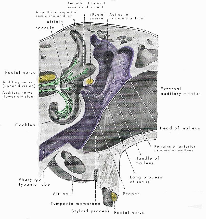
Figure 1. Tympanic cavity and adjacent parts.
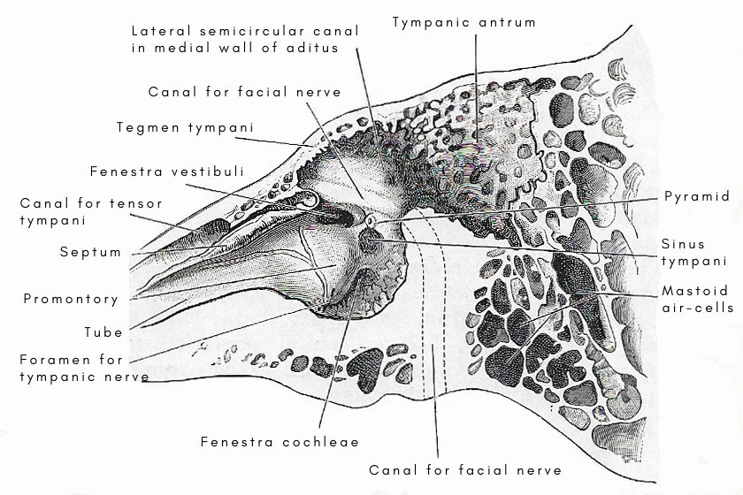
Figure 2. Vertical section through left ear.
The middle ear, also known as the tympanic cavity, is the air-filled cavity within the skull, located between the outer ear and the inner ear (Fig 1). The middle ear is linked to the pharynx (and therefore to the outside air) via the Eustachian tube and contains the three auditory ossicles, which transmit sound vibrations from the outer ear, via the tympanic membrane (eardrum), to the inner ear, via the oval window.
The middle ear is lined with mucous membrane and is filled with air. It communicates anteriorly with the pharynx through the pharyngo-tympanic tube, and posteriorly with the tympanic antrum and the mastoid air-cells, which are small cavities in the temporal bone.
Besides air, the middle ear contains: (1) the auditory ossicles – malleus, incus, and stapes; (2) two muscles – stapedius and tensor tympani; (3) the tympanic plexus of nerves and the chorda tympani nerve.
The vertical and antero-posterior diameters of the cavity are each about 15 millimeters. Its width, from side to side, is only about 6 millimeters above and 4 millimeters below, and is even less in the center because its lateral and medial walls both bulge into the cavity.
The part of the cavity above the level of the tympanic membrane is called the epitympanic recess.
The middle ear has a roof, a floor, and four walls – anterior, posterior, lateral, and medial.
Roof
The roof is a thin plate of bone – the tegmen tympani – which separates the cavity from the middle cranial fossa. In chronic inflammatory conditions of the middle ear, there is the risk that an extension of the inflammatory process through the tegmen may reach the meninges of the brain.
Floor
Th floor is narrow. It is a thin osseous lamina interposed between the tympanum and the jugular fossa, which is occupied by the upper bulb of the internal jugular vein, and an extension of an inflammatory condition of the middle ear through the bone may lead to thrombosis (clotting).
Posterior wall
The posterior wall presents: (1) in its upper part, the opening or aditus, which leads from the epitympanic recess into the tympanic antrum; (2) lower down, close to the medial wall, a small, hollow, conical projection called the pyramid (Fig 2); and (3) lateral to the pyramid, the opening through which the chorda tympani nerve enters the tympanum. The pyramid is perforated on its summit, and the aperture leads into a canal which curves backward and then downward until it opens into the lower part of the canal for the facial nerve. The curved canal of the pyramid lodges the stapedius muscle, the delicate tendon of which enters the tympanic cavity through the aperture on the summit of the pyramid.
Anterior wall
The anterior wall is narrow because the medial and lateral walls converge anteriorly. Its upper part is occupied by the opening of the tensor tympani canal; its middle part by the orifice of the pharyngo-tympanic tube; and the lowest part, which merges into the floor, is a lamina of bone which separates the tympanic cavity from the carotid canal. The septum between the tube and the tensor tympani canal is prolonged backward along the medial wall of the cavity as a shelf called the processus cochleariformis, and its end is molded to form a pulley round which the tendon of the tensor typani muscle turns abruptly in a lateral direction toward the malleus.
Medial wall
The medial wall intervenes between the middle ear and the inner ear. On it, there are eminences, depressions, and apertures. The anterior and larger part of the wall bulges into the cavity., and forms the very evident elevation of the promontory. Above the posterior part of the promontory is a depression leading to an oval aperture – the fenestra vestibuli. In the macerated bone, the fenestra opens into the vestibule of the inner ear; but, in the recent state, it is closed by the footpiece of the stapes – the most medial of the auditory ossicles. The pyramid, on the posterior wall, is immediately behind the fenestra vestibuli. Above the fenestra vestibuli, near the roof, there is an antero-posterior ridge produced by the canal for the facial nerve bulging into the tympanum. The wall of the canal is so thin the the facial nerve can be seen in it, Below and behind the promontory is a small, round hole – the fenestra cochleae. In the macerated bone, the fenestra leads into the cavity of the cochlea of the inner ear; but, in the recent state, it is closed by the secondary tympanic membrane.
Lateral wall
The lateral wall of the middle ear is formed, for the most part, by the tympanic membrane, but, above the membrane, the lateral wall of the epitympanic recess is formed by the squamous part of the temporal bone.

