
phalange
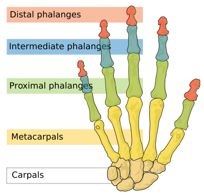
The phalanges and other bones of the hand.
A phalange is any of the bones in the fingers and toes. In humans, there are 14 phalanges in each hand and foot, 2 in each thumb and big toe, and 3 in the remaining digits. They are connected to the metacarpals in the hand and the metatarsals in the foot.
Osteology of phalanges of hand (Gray's Anatomy, 1918)
The phalanges of the hand (phalanges digitorum manus) are 14 in number, three for each finger, and two for the thumb. Each consists of a body and two extremities. The body tapers from above downward, is convex posteriorly, concave in front from above downward, flat from side to side; its sides are marked by rough areas which give attachment to the fibrous sheaths of the flexor tendons. The proximal extremities of the bones of the first row present oval, concave articular surfaces, broader from side to side than from before backward. The proximal extremity of each of the bones of the second and third rows presents a double concavity separated by a median ridge. The distal extremities are smaller than the proximal, and each ends in two condyles separated by a shallow groove; the articular surface extends farther on the volar than on the dorsal surface, a condition best marked in the bones of the first row.
The ungual phalanges are convex on their dorsal and flat
on their volar surfaces; they are recognized by their small size, and by
a roughened, elevated surface of a horseshoe form on the volar surface of
the distal extremity of each which serves to support the sensitive pulp
of the finger.
Articulations
In the four fingers the phalanges of the first row articulate with those of the second row and with the metacarpals; the phalanges of the second row with those of the first and third rows, and the ungual phalanges with those of the second row. In the thumb, which has only two phalanges, the first phalanx articulates by its proximal extremity with the metacarpal bone and by its distal with the ungual phalanx.
Ossification of the bones of the hand
The carpal bones are each ossified from a single center, and ossification proceeds in the following order: in the capitate and hamate, during the first year, the former preceding the latter; in the triangular, during the third year; in the lunate and greater multangular, during the fifth year, the former preceding the latter; in the navicular, during the sixth year; in the lesser multangular, during the eighth year; and in the pisiform, about the twelfth year.
Occasionally an additional bone, the os centrale, is found on the back of the carpus, lying between the navicular, lesser multangular, and capitate. During the second month of fetal life it is represented by a small cartilaginous nodule, which usually fuses with the cartilaginous navicular. Sometimes the styloid process of the third metacarpal is detached and forms an additional ossicle.
The metacarpal bones are each ossified from two centers: one for the body and one for the distal extremity of each of the second, third, fourth, and fifth bones; one for the body and one for the base of the first metacarpal bone. The first metacarpal bone is therefore ossified in the same manner as the phalanges, and this has led some anatomists to regard the thumb as being made up of three phalanges, and not of a metacarpal bone and two phalanges. Ossification starts in the middle of the body about the eighth or ninth week of fetal life, the centers for the second and third metacarpals being the first, and that for the first metacarpal, the last, to appear; about the third year the distal extremities of the metacarpals of the fingers, and the base of the metacarpal of the thumb, begin to ossify; they unite with the bodies about the twentieth year.
The phalanges are each ossified from two centers: one for the body, and one for the proximal extremity. Ossification begins in the body, about the eighth week of fetal life. Ossification of the proximal extremity starts in the bones of the first row between the third and fourth years, and a year later in those of the second and third rows. The two centers become united in each row between the eighteenth and twentieth years.
In the ungual phalanges the centers for the bodies appear at the distal extremities of the phalanges, instead of at the middle of the bodies, as in the other phalanges. Moreover, of all the bones of the hand, the ungual phalanges are the first to ossify.
Osteology of phalanges of foot (Gray's Anatomy, 1918)
The phalanges of the foot correspond, in number and general arrangement, with those of the hand; there are two in the great toe, and three in each of the other toes. They differ from them, however, in their size, the bodies being much reduced in length, and, especially in the first row, laterally compressed.
First row
The body of each is compressed from side to side, convex above, concave below. The base is concave; and the head presents a trochlear surface for articulation with the second phalanx.
Second row
The phalanges of the second row are remarkably small and short, but rather broader than those of the first row. The ungual phalanges, in form, resemble those of the fingers; but they are smaller and are flattened from above downward; each presents a broad base for articulation with the corresponding bone of the second row, and an expanded distal extremity for the support of the nail and end of the toe.
Articulations
In the second, third, fourth, and fifth toes the phalanges of the first row articulate behind with the metatarsal bones, and in front with the second phalanges, which in their turn articulate with the first and third: the ungual phalanges articulate with the second.
Ossification of the bones of the foot
The tarsal bones are each ossified from a single center, excepting the calcaneus, which has an epiphysis for its posterior extremity. The centers make their appearance in the following order: calcaneus at the sixth month of fetal life; talus, about the seventh month; cuboid, at the ninth month; third cuneiform, during the first year; first cuneiform, in the third year; second cuneiform and navicular, in the fourth year. The epiphysis for the posterior extremity of the calcaneus appears at the tenth year, and unites with the rest of the bone soon after puberty. The posterior process of the talus is sometimes ossified from a separate center, and may remain distinct from the main mass of the bone, when it is named the os trigonum.
The metatarsal bones are each ossified from two centers: one for the body, and one for the head, of the second, third, fourth, and fifth metatarsals; one for the body, and one for the base, of the first metatarsal. Ossification commences in the center of the body about the ninth week, and extends toward either extremity. The center for the base of the first metatarsal appears about the third year; the centers for the heads of the other bones between the fifth and eighth years; they join the bodies between the 18th and 20th years.
The phalanges are each ossified from two centers: one for the body, and one for the base. The center for the body appears about the tenth week, that for the base between the fourth and tenth years; it joins the body about the eighteenth year.

