aorta

Figure 1. Anatomy of the aorta.
Figure 2. The aortic valve and other major valves of the heart.
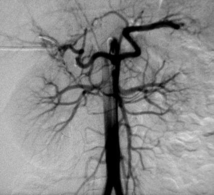
Figure 3. Abdominal aortography.
The aorta is the main artery of the body from which all others derive. It marks the beginning of the general or systemic circulation – as distinct from the pulmonary circulation. The aorta arises from the summit of the left ventricle (the main pumping chamber) of the heart, arches over the top of the heart, and descends in front of the backbone. It gives off large and small branches and finally divides to form the right and left iliac arteries in the legs.
Anatomically, the aorta is traditionally divided into the ascending aorta, the aortic arch, and the descending aorta. The short ascending portion runs up to the left of the superior vena cava. The aortic arch curves horizontally backward in front of the lower part of the trachea and over the left bronchus. The descending aorta runs down the spinal column and subdivides into the thoracic aorta, which descends within the chest, and the abdominal aorta, which descends within the abdomen.
The aorta gives off branches that go to the head and neck, the arms, the legs, the major organs in the chest and abdomen, and the heart itself (see coronary arteries). It serves to supply them all with oxygenated blood. The aorta is the central conduit from the heart to the body (Figure 1).
Branches of the aorta
Three great vessels arise from the top of the aortic arch. On the right is the innominate artery, which divides into the right subclavian artery and the right common carotid artery for the head and neck. On the left there is no innominate, the left subclavian and carotid arising directly from the arch. Only the lower parts of these vessels lie within the thorax, the level of the innominate bifurcation being at the inner end of the clavicle; they are closely applied to the trachea. The corresponding veins lie more superficially and there is an innominate vein on both sides; the vessel equivalent to the common carotid is the internal jugular vein. The small coronary arteries spring from the very beginning of the aorta, immediately above the semilunar valves. There is a right and left vessel, and any disease of these impairs the circulation in the muscular wall of the heart.
The descending aorta gives off on each side the pairs of intercostal arteries that encircle the chest in the intercostal spaces; the corresponding veins do not enter the venae cavae directly but through intervening channels, the azygous veins.
The aortic arch forms a great sweep from the front of the chest to the back, with an upward convexity from which arise the main arteries of the head, neck, and arms. The concavity of the arch embraces the pulmonary arteries and veins and the bronchi, entering the roots of the lungs. There is also a little artery running the length of the back of the sternum, from above downwards. This is the internal mammary artery, a branch of the subclavian in the neck; it ends below by entering the sheath of the rectus muscle as the superior epigastric artery to anastomose (see anastomosis) with the inferior epigastric from below.
The aortic arch inclines to the left, so that the descending portion is to the left of the midline and nearer the left lung.
Aortic valve
The aortic valve is a valve in the heart, lying between the left ventricle and the aorta (Figure 2). It is a semilunar valve that prevents blood returning to the ventricle from the aorta. See also heart valves.
Layers of the aorta
The aorta has a thick wall, with three layers of muscle that allow the blood vessel to withstand the high pressure that is generated when the heart pumps blood to the body. The three layers are the tunica intima, tunica media, and the tunic adventitia. The intima is the inside layer that is in contact with the blood, the media is in the middle, and the adventitia is the outermost layer.
Disorders of the aorta
Like other arteries, the aorta can become narrowed as a result of atherosclerosis (deposits of fat on the vessel walls), which often causes high blood pressure (hypertension). There are also a number of specific aortic disorders, notably coarctation of the aorta (in which the aorta is abnormally narrow at birth) and aortitis (inflammation of the wall of the aorta).
Both atherosclerosis and aortitis cause an aortic aneurysm, which may require surgery to correct impaired blood flow and to remove the risk of rupture and fatal blood loss.
An aortic dissection is a tear in the wall of the aorta, due to any of a variety of causes.
Aortography
Aortography is an imaging procedure by which the aorta (the main artery leaving the heart) and its branches can be seen on X-ray film after injection of a contrast medium (a substance that is opaque to X-rays). Aortography is a type of arteriography.
Why aortography is done
Aortography is used to detect aortic aneurysm (ballooning of the aorta) and assess the severity of peripheral arterial disease before surgery.
How it is done
Contrast medium is usually injected into the aorta through a fine catheter inserted either into the femoral artery at the groin, or into the brachial artery on the inside of the elbow. In people with severe arterial disease, the major arteries may be blocked and the contrast medium may have to be injected through a hollow needle directly into the aorta within the lower abdomen.
Complications
There is a small risk of a reaction to the contrast medium. Damage to a vessel during puncture or catheterization can also occur.