
trachea
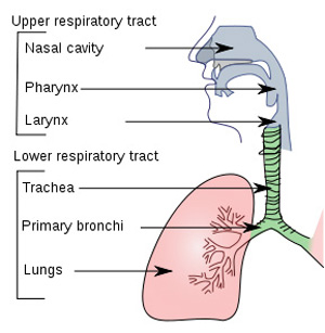
The respiratory tract with trachea shown in green.
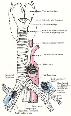
Larynx, trachea, and bronchi. The dotted line gives the outline of the thyroid gland.
The trachea, commonly called the windpipe, is the main airway to the lungs. It is a wide tube, 10–12.5 centimeters (4–5 inches) in length. It lies between the larynx and the main bronchi, i.e., from just below the Adam's apple, passing behind the notch of the sternum (breastbone) to behind the angle of the sternum. The trachea divides into the right and left bronchi at the level of the fifth thoracic vertebra, channeling air to the right or left lung.
The curved bars of hyaline cartilage in the tracheal wall provide support and keep the trachea from collapsing. The posterior soft tissue allows for expansion of the esophagus, which is immediately behind the trachea.
The mucous membrane that lines the trachea is ciliated pseudostratified columnar epithelium similar to that in the nasal cavity and nasopharynx. Goblet cells produce mucus that traps airborne particles and microorganisms, and the cilia propel the mucus upward, where it is either swallowed or expelled.
Thoracic portion of the trachea
The trachea begins in the neck at the lower end of the larynx, and about half of it is in the neck and half in the thorax. It enters the inlet of the thorax opposite the upper border of the manubrium sterni, and it terminates at the level of the lower border of the manubrium, opposite the interval between the third and fourth thoracic vertebrae, by dividing into a right and left bronchus; its thoracic part is therefore opposite the manubrium and wholly in the superior mediastinum. Its median axis is in the median plane except in its lower part, where it deviates slightly to the right. The trachea, being an elastic structure, elongates during inspiration; and its bifurcation may thus descend to the level of the body of the fifth or even the sixth thoracic vertebra.
Relations
Posteriorly, the trachea is in contact with the esophagus, which separates it from the vertebral column; and the left recurrent laryngeal nerve ascends in the groove between its left border and the esophagus.
Anteriorly, its lower part is crossed by the anterior part of the arch of the aorta; at a higher level, it is related to the innominate artery and left common carotid artery, with the left innominate vein and the remains of the thymus in front of them.
On the right side, it is in relation with the right pleura and lung, the right vagus nerve, and the arch of the azygos vein.
The upper part of its left side is related to the left common carotid artery and subclavian artery and the phrenic and vagus nerves, and the lower part to the arch of the aorta; and they separate it from the left pleura and lung.
Disorders of trachea
One of the most common disorders of the trachea is tracheitis (see below), which is usually caused by infection (especially by a virus) and is often associated with bronchitis and laryngitis. Obstruction of the trachea by an inhaled object is rare because the narrowest part of the upper respiratory tract is the larynx, and any objects that pass through there usually also continue through the trachea. However, the trachea may become obstructed by a tumor or narrowed as a result of scarring from a tracheostomy tube inserted to create an artificial airway through the front of the neck.
Rarely a congenital malformation, called a tracheoesophageal- fistula (see below) occurs in which an abnormal channel forms between the trachea and the esophagus immediately behind it.
Tracheitis
Inflammation of the trachea, usually caused by a viral infection. It is aggravated by inhaled fumes and especially by tobacco smoke, and often occurs with laryngitis and bronchitis, when it is known as laryngotracheobronchitis. Symptoms include a painful dry cough and hoarseness. In most cases, no treatment is needed.
Tracheoesophageal fistula
A rare birth defect in which an abnormal passage connects the trachea with the esophagus. In the most common form of this type of fistula, the lower end of the esophagus connects with the trachea, and the upper end of the esophagus is underdeveloped, forming a blind-ended pouch.

