
esophagus
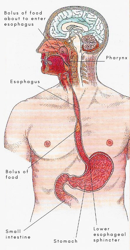
The esophagus is a collapsible muscular tube, about 25 cm (10 inches) long, that serves as a passageway between the pharynx and the stomach. It forms part of the gastrointestinal tract. Food passes down the esophagus by gravity and peristalsis.
As it descends, the esophagus is posterior to the trachea and anterior to the spinal column. It passes through an opening in the diaphragm, called the esophageal hiatus, and then empties into the stomach. The mucosa has glands that secrete mucus to keep the lining moist and well lubricated to ease the passage of food. Upper and lower esophageal sphincters control the movement of food into and out of the esophagus. The lower esophageal sphincter is sometimes called the cardiac sphincter and resides at the esophagogastric junction.
Further anatomy of the esophagus
the esophagus begins in the neck, where it is continuous with the lower part of the pharynx. It descends through the lower part of the neck behind the trachea, and enters the thorax at the inlet; it then descends through the superior and posterior mediastina, and leaves the thorax, opposite the seventh left cartilage about an inch from the median plane, at the level of the ninth thoracic spine, by passing through esophageal orifice of the diaphragm in the abdomen, where it joins the stomach almost at once. As it enters the thorax it is slightly to the left of the median plane, and trends more to the left as it descends, till, about the level of the seventh thoracic vertebra, it begins to pass from the medial side of the descending aorta on to the front of it; and, since the aorta, as it descends, inclines from the the left side of the vertebral bodies toward the median plane, the esophagus is even more to the left than the aorta before it pierces the diaphragm. Note that the esophagus is closely related to the aorta – first on its right side, and then in front of it.
Posterior relations
The vertebral column is behind the esophagus in the whole of its course with certain structures intervening; in the superior mediastinum, the longus cervicis muscles; in the upper part of the posterior mediastinum, upper five right aortic intercostal arteries, the thoracic duct, the aygos vein, and the hemiazygos veins; and in the upper part, the descending aorta.
Anterior relations
Anterior to the esophagus, in the superior mediastinum, are the trachea, the left recurrent laryngeal nerve, and the structures which lie still further forward. As it passes from the superior to the posterior mediastinum, its anterior relations are first the start of the left bronchus and the right pulmonary artery. In the posterior mediastinum, the esophagus is separated from the left atrium by the posterior wall of the pericardium; and, at a lower level, the diaphragm is in front of it.
Right relations
In the superior mediastinum, the esophagus is in relation with the right pleura and lung, and with the arch of the azygos vein; and, in the posterior mediastinum, with the right pleura and lung,until it passes forward and toward the left on to the front of the descending aorta.
Left relations
In the superior mediastinum, the esophagus is in relation with the thoracic duct, the left subclavian artery, the left pleura and lung, and the arch of the aorta. From the fifth to the seventh thoracic vertebrae, its left lateral relation is the descending aorta; and its lower part, which lies in front of the descending aorta, is in relation with the left pleura and lung.
Functions of the esophagus
Disorders
Disorders and diseases of the esophagus include reflux esophagitis or heartburn, ulcer, stricture, and cancer.

