
trigeminal nerve
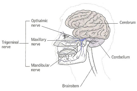
The trigeminal nerve is a paired nerve arising from the brainstem and supplied the face. Each one of the pairs supplies one side of the face. The trigeminal nerve splits into three main branches. The opthalmic nerve supplies most of the scalp, the upper eyelid, and the cornea; the maxillary nerve supplies the upper jaw; and the mandibular nerve supplies the tongue, lower jaw, and jaw muscles.
The trigeminal nerve is the fifth cranial nerve. It is the thickest of the cranial nerves, and has a wide distribution. Its motor fibers supply the four muscles of mastication (chewing) and four other muscles – tensor palati, tensor tympani, mylo-hyoid, and anterior belly of the digastric. Its sensory fibers supply: (1) part of the dura mater; (2) the eyeball (see eye) and other contents of the orbit; (3) the skin of face and anterior half of the head; (4) the mucous membranes of the air sinuses, nose, mouth, and anterior two-thirds of the tongue; (5) the teeth; (6) the mandibular joint (see mandible).
Like a spinal nerve, the trigeminal nerve has two roots – a sensory and a motor – and there is a ganglion on the sensory root. The sensory fibers arise in the ganglion. Their central branches emerge to form the three great divisions (hence the name "trigeminal") of the trigeminal nerve:
The mandibular division enters a canal in the lower jaw. The ophthalmic division enters the back of the orbit. The maxillary division runs in a canal between the orbital floor and the roof of the maxilla to emerge on the front of the cheek.
The motor root of the trigeminal arises in the pons and eventually joins the mandibular nerve as it leaves the skull.
The sensory and motor roots of the trigeminal nerve are enclosed in an adherent sheath of pia mater and a loose sheath of arachnoid mater that contains cerebrospinal fluid. Traced from the pons toward the ganglion, the two roots cross the medial part of the upper border of the petrous temporal bone through an aperture in the dura mater.
Trigeminal ganglion
The trigeminal ganglion lies in a shallow depression on the front of the petrous temporal bone near its apex and on the suture (or gap) between that part of the petrous temporal and the greater wing of the sphenoid bone.
It occupies a space between the outer and inner layers of the dura mater called the cavum trigeminale, but is enclosed in a pocket or diverticulum of the inner layer which is prolonged over it from the lips of the aperture that transmits the roots. There are therefore two layers of dura mater above it and two below it; but the two below it are fused together. The motor root passes below the ganglion to join the mandibular nerve. The internal carotid artery is first below it in the carotid canal, and then medial to it in the cavernous sinus; and the sympathetic plexus around the artery is connected with the ganglion by communicating filaments. The ganglion is crescentic in outline, the concavity being directed upward and backward. The sensory root emerges from the concave border; and the three great divisions emerge from the convex border.
Sensory root of the trigeminal nerve
The sensory root, on emerging from the ganglion, is at first wide and plexiform and lies in the depression on the front of the petrous temporal bone. Then, narrowing, it crosses the upper border of the petrous temporal below the attached margin of the tentorium, and passes horizontally backward and slightly medially to the pons.
Motor root of the trigeminal nerve
This root is much smaller than the sensory root. It issues from the pons, runs along the sensory root, at first on its medial side and then below it, and descends under cover of the ganglion and mandibular nerve to the foramen ovale. There, it joins the mandibular nerve, which is therefore the only one of the three divisions of the trigeminal nerve that contains motor fibers.
Trigeminal nerve neuralgia
This is a disorder of the trigeminal nerve in which brief episodes of severe, stabbing pain affect the cheeks, lips, gums, or chin on one side of the face. The disorder usually occurs in people over the age of 50. Pain may come in bouts that last for weeks at a time. The cause is uncertain, and pain is often brought on by touching the face, eating, drinking, or talking.

