cervical vertebra
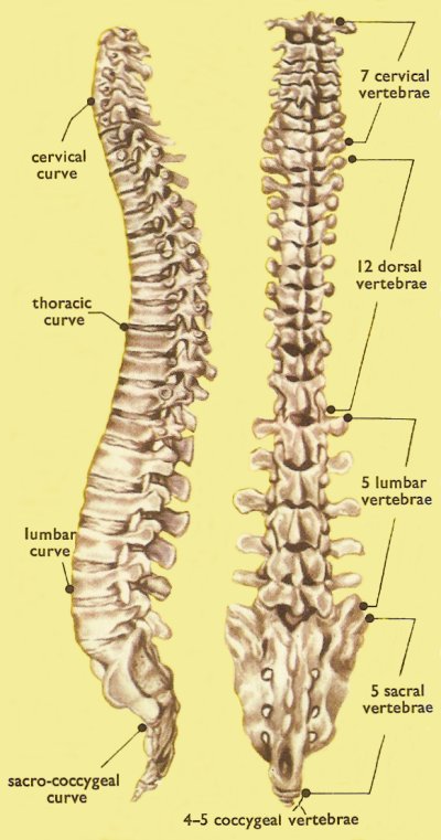
Figure 1. Spinal column, lateral and posterior views.
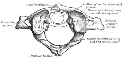
Figure 2. Atlas (C1), Gray's Anatomy (1918).
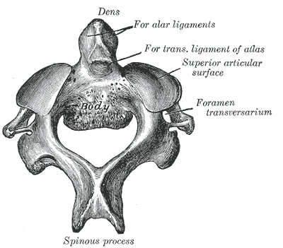
Figure 3. Axis (C2), Gray's Anatomy (1918).
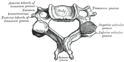
Figure 4. Typical cervical vertebra (C3-C6), Gray's Anatomy (1918).
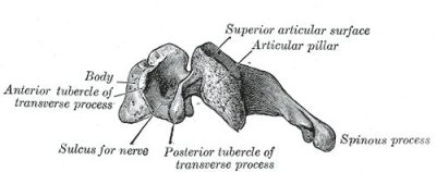
Figure 5. Typical cervical vertebra (side view), Gray's Anatomy (1918).
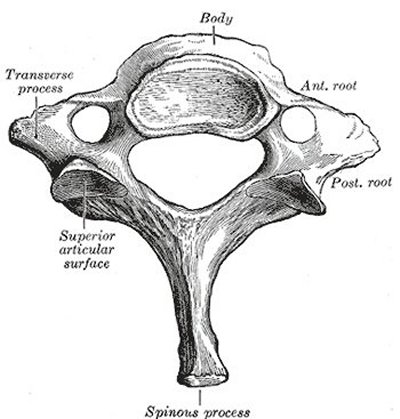
Figure 6. Seventh cervical vertebra, Gray's Anatomy (1918).
A cervical vertebra is any of the vertebra in the cervical (neck) region of the spinal column. The cervical vertebra are the smallest vertebra in the spine, reflective of the fact that they support the least load.
In humans, and almost all other mammals, there are seven cervical vertebra, which are labeled C1 to C7. Anatomists divide them into two regions: the upper cervical region (C1 and C2), and the lower cervical region (C3 through C7). Three cervical vertebra have a unique anatomical structure and have been given special names. C1 is called the atlas, C2 the axis, and C7 the vertebra prominens.
Only the cervical vertebrae have three openings or foramina – the vertebral foramina and two transverse foramina. A characteristic feature of the vertebrae C2 to C6 is a projection known as the bifid spinous process. C7 has a prominent nonbifid spinous process that can be felt at the base of the neck.
Atlas (C1)
The atlas, or first cervical vertebra (Figure 2), is so named because it supports the globe of the skull. Its appearance is quite different from the other spinal vertebrae. Most notably it has no body or spinous process, but instead consists of a ring of bone made up of two lateral masses joined at the front and back by the anterior arch and the posterior arch.
Axis (C2)
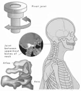
The axis (Figure 3) is the second cervical vertebra. Its most distinctive feature is a blunt tooth-like process, called the dens (Latin for "tooth") or odontoid process, which projects upward. The dens provides a kind of pivot and collar allowing the head and atlas to rotate around the dens.
 |
Typical cervical vertebra
Note: the following paragraph is adapted from Gray's Anatomy, 1918 ed.
The body is small, and broader from side to side than from front to back Figures 3 and 4). The anterior and posterior surfaces are flattened and of equal depth; the former is placed on a lower level than the latter, and its inner border extends downward, so as to overlap the upper and forepart of the vertebra below. The upper surface is concave transversely, and presents a projecting lip on either side; the lower surface is concave from front to back, convex from side to side, and presents laterally shallow concavities which receive the corresponding projecting lips of the subjacent vertebra. The pedicles are directed lateralward and backward, and are attached to the body midway between its upper and lower borders, so that the superior vertebral notch is as deep as the inferior, but it is, at the same time, narrower. The laminae are narrow, and thinner above than below; the vertebral foramen is large, and of a triangular form. The spinous process is short and bifid, the two divisions being often of unequal size. The superior and inferior articular processes on either side are fused to form an articular pillar, which projects lateralward from the junction of the pedicle and lamina. The articular facets are flat and of an oval form: the superior look backward, upward, and slightly medialward: the inferior forward, downward, and slightly lateralward. The transverse processes are each pierced by the foramen transversarium, which, in the upper six vertebrae, gives passage to the vertebral artery and vein and a plexus of sympathetic nerves. Each process consists of an anterior and a posterior part. The anterior portion is the homologue of the rib in the thoracic region, and is therefore named the costal process or costal element: it arises from the side of the body, is directed lateralward in front of the foramen, and ends in a tubercle, the anterior tubercle. The posterior part, the true transverse process, springs from the vertebral arch behind the foramen, and is directed forward and lateralward; it ends in a flattened vertical tubercle, the posterior tubercle. These two parts are joined, outside the foramen, by a bar of bone which exhibits a deep sulcus on its upper surface for the passage of the corresponding spinal nerve.
Vertebra prominens (C7)
Note: the following paragraph is adapted from Gray's Anatomy, 1918 ed.
The most distinctive feature of the C7 vertebra (Figure 6) is a long and prominent spinous process. This process is thick, nearly horizontal, not bifurcated, but terminating in a tubercle to which the lower end of the ligamentum nuchae is attached. The transverse processes are of considerable size, their posterior roots are large and prominent, while the anterior are small and faintly marked; the upper surface of each has usually a shallow sulcus for the eighth spinal nerve, and its extremity seldom presents more than a trace of bifurcation. The foramen transversarium may be as large as that in the other cervical vertebrae, but is generally smaller on one or both sides; occasionally it is double, sometimes it is absent. On the left side it occasionally gives passage to the vertebral artery; more frequently the vertebral vein traverses it on both sides; but the usual arrangement is for both artery and vein to pass in front of the transverse process, and not through the foramen. Sometimes the anterior root of the transverse process attains a large size and exists as a separate bone, which is known as a cervical rib.
Cervical vertebra in other animals
All mammals, however long or short their necks may be, have seven cervical vertebrae, except for manatees and two-toed sloths, each of which have six, and three-toed sloths, which have nine. The variation in number is much greater among mammals in the case of the vertebrae in other regions of the spinal column. And across all types of vertebrates, there is much variety with regard to both the number and structure of the vertebrae.


