
elbow
Right elbow joint.
Elbow joint.
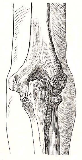
Relation of bones of the elbow to the surface. Dorsal view; elbow fully extended.
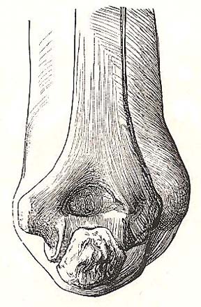
Relation of bones of the elbow to the surface. Dorsal view; elbow fully extended.
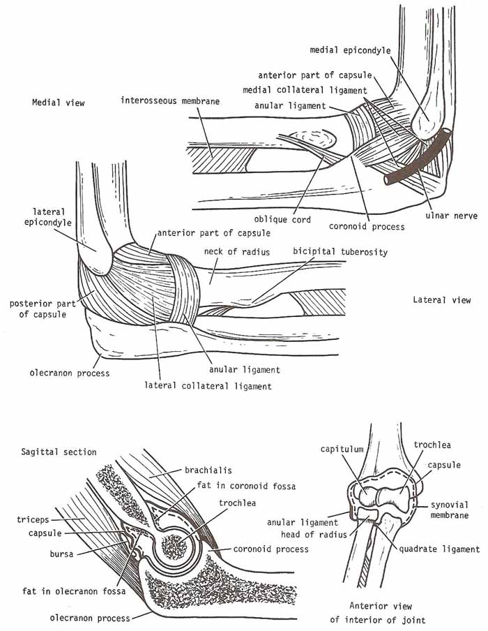
The elbow is the hinge joint in the arm that occurs between the trochlea and capitulum the humerus (the upper arm bone) and the trochlear notch of the ulna and the head of the radius (the bones of the forearm). The ulna forms the part of the joint that allows the hinge motion of the elbow. Part of the radius bone of the forearm (the head of the radius) sits against the humerus and turns to allow the forearm to turn so that the palm is up (pronation) or down (supination).
The elbow joint is very stable because of the wrench shape of the trochlear notch of the ulna, which fits around the pulley-shaped trochlea of the humerus. The joint is also strengthened by strong medial and lateral collateral ligaments.
The surface of the elbow joint is covered by hyaline cartilage that protects and cushions the joints. The large muscle in the back of the arm, the triceps, attaches to the point of the ulna (called the olecranon). When this muscle contracts, it straightens out the elbow. The biceps muscle in the front of the arm, when contracted, bends the elbow.
Capsule of the elbow joint
Anteriorly, the capsule is attached to the humerus along the upper margins of the coronoid and radial fossae and to the front of the medial and lateral epicondyles. Below, it is attached to the margin of the coronoid process of the ulna and to the annular ligament, which surrounds the head of the radius.
Posteriorly, the capsule is attached above to the margins of the olecranon fossa of the humerus. Below, it is attached to the upper margin and sides of the olecranon process of the ulna and to the annular ligament.
Ligaments of the elbow joint
The lateral ligament is triangular in shape and is attached by its apex to the lateral epicondyle of the humerus, and by its base to the upper margin of the annular ligament.
The medial ligament is also triangular in shape and consists principally of three strong bands: (1) The anterior band, which passes from the medial epicondyle of the humerus to the medial margin of the coronoid process. (2) The posterior band, which passes from the medial epicondyle of the humerus to the medial side of the olecranon. (3) The transverse band, which passes between the ulnar attachments of the two preceding bands.
Synovial membrane
The synovial membrane of the elbow joint lines the capsule and covers the floors of the coronoid, radial, and olecranon fossae; it is continuous below with the synovial membrane of the superior radioulnar joint.
Movements of the elbow joint
The elbow joint is capable of flexion and extension. Flexion is limited by the anterior surfaces of the forearm and arm coming into contact. Extension is checked by the tension of the anterior ligament and the brachialis muscle. Flexion is performed by the brachialis, biceps brachii, brachioradialis, and pronator teres muscles. Extension is performed by the triceps and anconeus muscles.
The long axis of the extended forearm lies at an angle to the long axis of the arm. This angle, which opens laterally, is called the carrying angle and is about 170° in the male and 167° in the female. The angle disappears when the elbow is fully flexed.
Important anatomical relations
Anteriorly: The brachialis, the tendon of the biceps, the median nerve, and the brachial artery.
Posteriorly: The triceps muscle, a small bursa intervening.
Medially: The ulnar nerve passes behind the medial epicondyle and crosses the medial ligament of the joint.
Laterally: The common extensor tendon and the supinator.
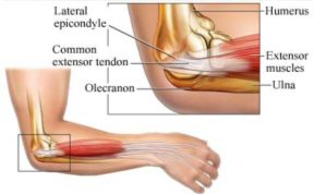
Tennis elbow
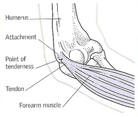 |
| The site of tennis elbow. Pulling
of the forearm muscles at the point where they attach to the humerus
causes tenderness on the outer side of the elbow.
|
Tennis elbow is pain and tenderness on the outside of the elbow and in the back of the forearm. Also called lateral epicondylitis, it is caused by inflammation of the tendon that attaches the muscles that straighten the fingers and wrist to the humerus (the upper-arm bone).
The treatment of tennis elbow consists of resting the arm, applying ice-packs, and taking analgesic drugs (pain-killers), or non-steroidal anti-inflammatory drugs (NSAIDs). Ultrasound treatment, injection of a corticosteroid drug, or surgery are sometimes needed.

