
humerus
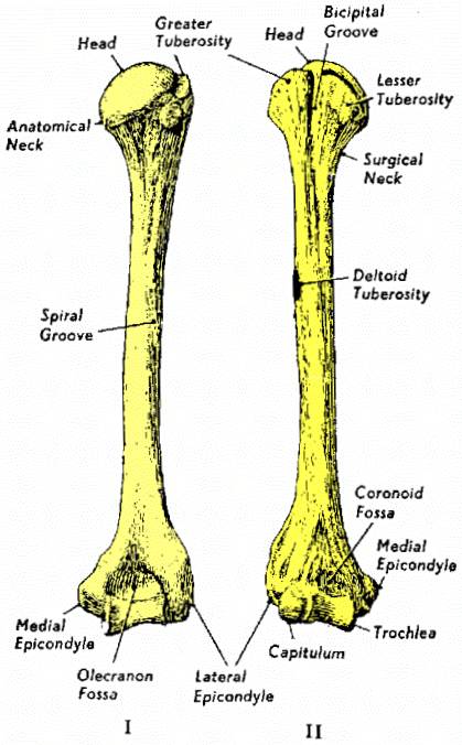
Posterior (I) and anterior
(II) view of the right humerus.
Illustration: Gray's Anatomy.
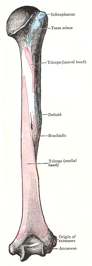
Back of the humerus with muscular attachments mapped out.
The humerus is the long bone of the upper arm of humans and other tetrapod vertebrates.
The humerus extends from the scapula, or
shoulder-blade, where it articulates at the glenoid
cavity, to the elbow. A notch, called
the olecranon cavity, on the posterior roughened end of
the humerus, provides the point of articulation for the ulna,
one of the bones of the forearm.
Detailed anatomy
The head of the humerus at the proximal end forms slightly less than a sphere. Its articular surface covered by hyaline cartilage faces upwards, inwards and slightly backwards. The curvature of the head is not reciprocal to that of the glenoid cavity. This means that only a small area of the head is in contact with the scapula at any one time.
The anatomical neck is the slight constriction just behind the head. The surgical neck is the first narrowing on the shaft below the expanded proximal end.
The lesser tubercle projects forwards from just beyond the anterior part of the anatomical neck. The greater tubercle is the most lateral part of the proximal humerus, producing the rounded contour of the shoulder.
The shaft of the humerus is roughly cylindrical proximally, becoming triangular distally. About mid-way down the shaft on the lateral aspect is a rough area, the deltoid tuberosity. Curving around the posterior aspect of the upper half of the shaft is the spiral groove.
The distal expanded end of the humerus carries articular surfaces for the ulna and radius and attachments (the epicondyles) for the forearm muscles. Laterally and anteriorly the capitulum forms a small hemisphere, articulating with the discoid radial head. Anteriorly in the middle third of the distal expansion the pulley-shaped trochlea articulates with the trochlear notch of the ulna. The medial third is formed into the medial epicondyle, the common origin of the flexors of the forearm.
Depressions are present in the anterior aspect, above the trochlea and capitulum for the ulna (coronoid fossa) and above the capitulum for the radius (radial fossa). Similarly a depression is present above the trochlea on the posterior aspect for the olecranon process of the ulna (olecranon fossa).
Fractures of the humerus
Fractures of the humerus may affect the upper end, the shaft, or the lower end of the bone.
The part of the humerus most commonly fractured is the neck of the bone (at the upper end, just below the head of the humerus). This type of fracture most often occurs in elderly people. Fractures of the shaft usually affect the middle third of the bone. Such fractures occur in adults of all ages but are uncommon in children. Supracondylar fractures (at the lower end of the bone) occur most commonly in children.
An X-ray is performed if any fracture of the humerus is suspected.
A fracture of the neck of the bone requires only a sling to keep the bone immobilized: a fracture of the shaft or of the lower end of the bone usually needs to be put in a plaster cast. Most fractures of the humerus heal in six to eight weeks.
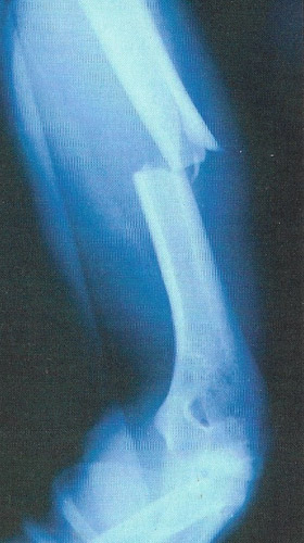 |
| A fracture of the humerus is shown in this X-ray. The broken bone ends are displaced and willneed to be manipulated back into position, then immobilized in a plaster cast. |
Complications
Damage to the radial nerve may occur when the shaft of the humerus is fractured. In severe cases, this may result in wrist-drop. Fracture of the humerus may be accompanied by damage to the brachial artery. If such damage is undetected, circulation to the arm may be impaired, resulting in Volkmann's contracture (a deformity of the forearm and hand due to muscle damage caused by insufficient blood supply).
Some supracondylar fractures fail to mend properly – despite all remedial efforts and tractions – resulting in deformity of the elbow and an increased risk that osteoarthritis will eventually develop.

