
utricle
The utricle, also called the utriculus, is a dilated portion of the membranous labyrinth within the inner ear. It occupies the central part of the bony labyrinth, with the cochlea in front and the semicircular canals, which arise from the utricle, behind.
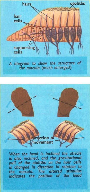
Within the utricle a small projection, known as the macula, is raised from the epithelium inside the membranous labyrinth. It consists of a group of supporting cells, among which are arranged a number of hair cells. Each hair cell has a fine hair process, which extends from the end of the cell into the cavity of the utricle. These hairs are embedded in a jelly-like material, which contains a large number of calcareous bodies known as otoliths.
The hair cells of the macula are attached to the nervous system by fibers of the vestibular division of the auditory nerve, each cell being served by one fiber.
Next to the utricle is a very similar organ called the saccule. It is also is provided with a macula, like the utricle.
Function of the utricle
The otoliths in the utricle are subject to the action of gravity, and since they are in contact with the hairs of the hair cells the gravitational pull upon them is transmitted to the macula. Furthermore, since gravity always pulls the otoliths towards the center of the Earth, a change in the position of the head, and therefore of the utricle, alters the direction in which the otoliths pull in relation to the macula. As a result the stimulus to the hair cells changes, and a different sequence of nerve impulses passes along the vestibular division to the brain.
In this way the utricle continually reports the position of the head. This information, when associated with information from the muscles, is enough to indicate the position of the body as a whole. Thus even a blindfold person knows the exact position in space of each part of her body. See also vestibular sense.

