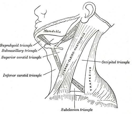
posterior triangle (of the neck)

The posterior triangle is made up of the occipital and subclavian triangles.
The posterior triangle of the neck is the triangular space behind the sternomastoid, a muscle that passes obliquely across the neck, from the sternum and clavicle below, to the mastoid process and occipital bone above. The similar space in front of this muscle is called the anterior triangle.
Behind the posterior triangle is the anterior margin of the trapezius. The base of the triangle is formed by the middle third of the clavicle; its apex, by the occipital bone. The space is crossed, about 2.5 centimeters. above the clavicle, by the inferior belly of the omohyoid, which divides it into two smaller triangles, an upper or occipital, and a lower or subclavian.
The occipital triangle – the larger division of the posterior triangle – is bounded, in front, by the sternomastoid; behind, by the trapezius; and below, by the omohyoid. Its floor is formed from above downward by the splenius capitis, levator scapulae, and the scaleni medius and posterior. It is covered by skin, superficial and deep fascia, and by the platysma below. The accessory nerve is directed obliquely across the space from the sternomastoid, which it pierces, to the under surface of the trapezius; below, the supraclavicular nerves, transverse cervical vessels, and upper part of the brachial plexus cross the space. A chain of lymph glands is also found running along the posterior border of the sternomastoid, from the mastoid process to the root of the neck.
The subclavian triangle is bounded, above, by the inferior belly of the omohyoid; below, by the clavicle; its base is formed by the posterior border of the sternomastoid. Its floor is formed by the first rib with the first digitation of the serratus anterior. The size of the subclavian triangle varies with the extent of attachment of the clavicular portions of the sternomastoid and trapezius, and also with the height at which the omohyoid crosses the neck. Its height also varies according to the position of the arm, being diminished by raising the limb, on account of the ascent of the clavicle, and increased by drawing the arm downward, when that bone is depressed. This space is covered by the integument, the superficial and deep fasciae, and the platysma, and crossed by the supraclavicular nerves. Just above the level of the clavicle, the third portion of the subclavian artery curves laterally and downward from the lateral margin of the scalenus anteriorscalenus anterior, across the first rib, to the axilla, and this is the situation most commonly chosen for ligaturing the vessel. Sometimes this vessel rises as high as 4 cm above the clavicle; occasionally, it passes in front of the scalenus anterior, or pierces the fibers of that muscle. The subclavian vein lies behind the clavicle, and is not usually seen in this space; but in some cases it rises as high as the artery, and has even been seen to pass with that vessel behind the scalenus anterior. The brachial plexus of nerves lies above the artery, and in close contact with it. Passing transversely behind the clavicle are the transverse scapular vessels; and traversing its upper angle in the same direction, the transverse cervical artery and vein. The external jugular vein runs vertically downward behind the posterior border of the sternomastoid, to terminate in the subclavian vein; it receives the transverse cervical and transverse scapular veins, which form a plexus in front of the artery, and occasionally a small vein which crosses the clavicle from the cephalic. The small nerve to the subclavius also crosses this triangle about its middle, and some lymph glands are usually found in the space.


