
subclavian artery
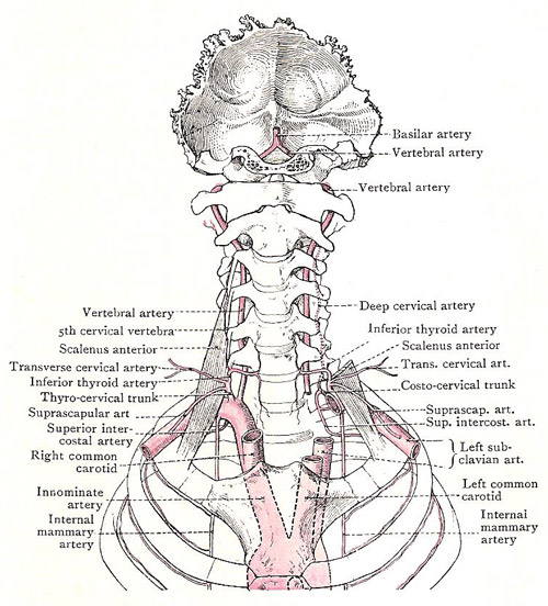
Subclavian arteries and their branches.
The subclavian artery is either of two arteries that distributes blood to the neck, thoracic (chest) wall, spinal cord, brain, meninges, and upper limbs.
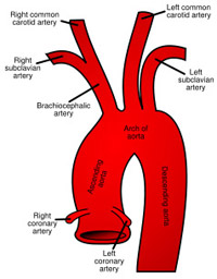
The right subclavian artery arises from the brachiocephalic artery, behind the right sternoclavicular joint. It passes upward and laterally as a gentle curve behind the scalenus anterior muscle, and at the outer border of the first rib becomes continuous with the axillary arch.
The left subclavian artery arises from the posterior part of the arch of the aorta, a little behind the left common carotid artery. It ascends vertically to the root of the neck, opposite the left sternoclavicular joint, and then arches laterally in a manner similar to that of the right subclavian artery. Like the common carotid artery, it has no branches on the thorax.
For descriptive purposes, the artery is divided into three parts. The first part extends from the origin of the vessel to the medial margin of the scalenus anterior; the second part lies behind that muscle; and the third part extends from its lateral border to the outer border of the first rib.
 |
| Origin of the subclavian arteries |
First part of the subclavian
Owing to the difference of origin, the relations of the first portion of the subclavian artery are not the same on the two sides of the body.
The first part of the right subclavian extends obliquely upwards and laterally, and at its termination at the medial margin of the scalenous anterior it has reached a point about half an inch above the level of the clavicle. It is placed very deeply. Anteriorly, it is covered by the skin, superficial fascia, platysma, deep fascia, and three muscular strata – the sternomastoid, sternohyoid, and sternothyroid. Two veins and some nerves are in front of it: at the medial margin of the scalenus anterior it is crossed by the internal jugular and vertebral vein. The nerves which cross in front of it are the vagus, a loop from the sympathetic (ansa subclavia), and, sometimes, cardiac branches of the vagus and sympathetic as they run into the thorax. The common carotid artery also is in front of it, near its origin.
The cervical plexus is both below and behind the artery, separated from it by a thin fibrous sheet called the suprapleural membrane. At the lower margin of the artery, the vagus gives off its recurrent laryngeal branch, which hooks round below it and ascends behind it.
The first part of the left subclavian ascends almost vertically from the aortic arch, and, when it reaches the root of the neck, it passes upwards and laterally to the medial margin of the scalenus anterior. The relations of the cervical part are slightly different from those on the right side. The same fascial and muscular layers, and the same nerves and veins, are in front of it. But in the rest of the neck, the veins and nerves of both sides trend to the right; on the left side, therefore, they are more or less parallel to the part of the subclavian artery that ascends behind the sternoclavicular joint. Three additional relations are established – the phrenic nerve and the thoracic duct descend in front of it near the scalenus anterior; and the left innominate vein is in front of it, behind the joint.
The left recurrent nerve hooks round the arch of the aorta, and lies to the medial side of the subclavian artery.
Second part
The second portion of the subclavian artery forms the highest part or summit of the arch, and rises above the level of the clavicle for half an inch to an inch.
In that part of its course the vessel has not so many superficial relations. Anteriorly, it is covered by (1) skin; (2) superficial fascia and platysma; (3) deep fascia; (4) clavicular head of the sternomastoid; (5) scalenus anterior. On the right side the phrenic nerve also is an anterior relation, but it is separated from the artery by the medial margin of the scalenus anterior. Posteriorly and inferiorly, the vessel is in relation with the pleura – the suprapleural membrane intervening.
Branches of the subclavian artery
Four branches spring from the subclavian trunk. Three take origin, as a general rule, from the first part of the artery, and one from the second part. They are: from the first part, the vertebral artery, thyrocervical trunk, and internal mammary artery, and from the second part the costocervical trunk.

