
stress testing
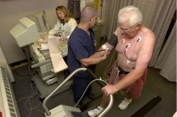
A patient walks on a stress test treadmill to check his heart's functioning.
Stress testing provides a doctor with information about how a person's heart works during physical stress. Some heart problems are easier to diagnose when the heart is working hard and beating fast. During a stress test, the subject exercises (walks or runs on a treadmill or pedal a bicycle) or is given a medicine to make the heart work harder while heart tests are performed.
During these tests, the heart is monitored using images or through dime-sized electrodes attached to the chest, arms, or legs. The subjectmay be asked to breathe into a special tube during the test. This allowa the doctor to see how well the subject is breathing.
A patient may have arthritis or another medical problem that prevents them from exercising during a stress test. If so,the doctor may administer a medicine that makes the heart work harder, as it would during exercise. This is called a pharmacological stress test.
Overview
Doctors usually use stress testing to help diagnose coronary artery disease (CAD) or to see how serious this disease is in those who are known to have it. It's sometimes used to assess other problems such as heart valve abnormalities or heart failure.
CAD occurs when the arteries that supply blood to the heart muscle (the coronary arteries) become hardened and narrowed with a material called plaque. Plaque is made up of fat, cholesterol, calcium, and other substances found in the blood. Plaque builds up on the insides of the arteries, narrowing them and restricting blood flow to your heart.
You may not have any signs or symptoms of CAD when your heart is at rest. But when your heart has to work harder during exercise, it needs more blood and oxygen, and narrowed arteries aren't able to supply enough blood for your heart to work well. Thus, the signs and symptoms may occur only during exercise.
A stress test can detect the following indications that your heart may not be getting enough blood during exercise.
During the stress test, if you can't exercise for as long as what's considered normal for someone your age, it may be a sign that not enough blood is flowing to your heart. But other factors besides CAD can prevent you from exercising long enough (for example, lung diseases, anemia, or poor general fitness).
Stress testing using imaging
Some stress tests take pictures of the heart when you exercise and when you're at rest. These imaging stress tests can show how well blood is flowing in the different parts of your heart and/or how well your heart squeezes out blood when it beats.
One type of imaging stress test involves echocardiography, which is a test that uses sound waves to create a moving picture of your heart. An echocardiogram stress test can show how well your heart's chambers and valves are working when your heart is under stress. The test can identify areas of poor blood flow to your heart, dead heart muscle tissue, and areas of the heart muscle wall that aren't contracting normally. These areas may have been damaged during a heart attack or may be getting too little blood.
Other imaging stress tests use a radioactive dye to create images of the blood flow to your heart. The dye is injected into your bloodstream before pictures are taken of your heart. The pictures show how much of the dye has reached various parts of your heart during exercise and at rest.
Tests that use a radioactive dye include a thallium or sestamibi stress test and a positron emission tomography (PET) stress test. The amount of radiation in the dye is safe and not a danger to you or those around you. However, if you're pregnant, you shouldn't have this test because of risks it might pose to your unborn child.
Some doctors may use magnetic resonance imaging (MRI) to take pictures of the heart when it's working hard. This test doesn't use a radioactive dye or sound waves. Instead, it uses radio waves and magnetic fields to create images that show blood flow in the heart and whether all parts of the heart wall are contracting strongly.
Imaging stress tests tend to be more accurate at detecting CAD than standard (nonimaging) stress tests. An imaging stress test may be done first if you:
Who needs stress testing?
You may need a stress test if you've had chest pains, shortness of breath, or other symptoms of limited blood flow to your heart. Imaging stress tests are particularly helpful in showing whether you have coronary artery disease (CAD) or a problem with one of the valves in your heart. (Heart valves are like doors that let blood flow between the heart's chambers and into the heart's arteries. So, like CAD, faulty heart valves can limit the amount of blood reaching your heart.)
If you've been diagnosed with CAD or recently had a heart attack, you may need stress testing to see whether you can tolerate an exercise program. The testing also can show whether treatments designed to improve blood flow in the heart's arteries are necessary and likely to help you. These treatments include angioplasty (with or without stents) and coronary artery bypass grafting. After having one of these treatments, your doctor may want you to have a stress test to see how well the treatment relieves your signs or symptoms of CAD.
You also may need a stress test if, during exercise, you feel faint, get a rapid heartbeat or a fluttering feeling in your chest, or have other symptoms of an arrhythmia (an irregular heartbeat). The stress test can detect an arrhythmia and show whether you need medicine or a pacemaker or implantable cardioverter defibrillator (ICD) to correct irregular heartbeats. It also can reveal the effectiveness of such devices.
You may need a stress test even if you don't have chest pain when you exercise, but just get short of breath. The test can help show whether a heart problem, rather than a lung problem or being out of shape, is causing your breathing problems. For such testing, you breathe into a special tube so a technician can measure the gases you breathe out.
Breathing into the special tube and monitoring of the heart as part of a stress test also is done to assess fitness before a heart transplant. Your doctor also may use this monitoring to figure out the best exercise plan for you after recovery from a heart attack.
Stress testing isn't routinely done to screen people for CAD. Usually you have to have symptoms of CAD before a doctor will recommend that you have a stress test. But your doctor may want to use a stress test to screen for CAD if you have diabetes, which increases your risk for developing CAD.
Before stress testing
Standard stress testing can often be done in a doctor's office. But imaging stress testing is usually done at a hospital. Be sure to wear athletic or other shoes in which you can exercise comfortably. You may be asked to wear comfortable clothes in which you can easily exercise, or you may be given a gown to wear during the test.
Your doctor may ask you not to eat or drink anything but water for a short time before the test. If you're diabetic, ask your doctor whether you need to adjust your medicines on the day of your test.
For some stress tests, you can't drink coffee or other caffeinated drinks for a day before the test. Certain over-the-counter or prescribed medicines also may interfere with some stress tests. Ask your doctor whether you can take all your medicines as usual and whether you need to avoid certain drinks or foods.
If you use an inhaler for asthma or other breathing problems, bring it to the test and be sure to let the doctor know that you use it.
During stress testing
During all types of stress testing, a technician will always be with you to closely monitor your health status.
Before you start the "stress" part of a stress test, a technician will put small sticky patches called electrodes on the skin of your chest, arms, and legs. To help an electrode stick to the skin, the technician may have to shave a patch of hair where the electrode will be attached.
The electrodes are connected to a machine that records the electrical activity of your heart. This recording, which is called an electrocardiogram (EKG), shows how fast your heart is beating and the heart's rhythm (steady or irregular). The machine also records the strength and timing of electrical signals as they pass through each part of your heart.
The technician will put a blood pressure cuff on your arm to monitor your blood pressure during the stress test. (The cuff will feel tight on your arm when it expands every few minutes.) In addition, you may be asked to breathe into a special tube so the gases you breathe out can be monitored.
After these preparations, you will exercise on a treadmill or stationary bicycle. If such exercise poses a problem for you, you may instead turn a crank with your arms. During the test, the exercise level will get harder. But you can stop whenever you feel the exercise is too much for you.
If you can't exercise, a technician will inject a medicine into a vein in your arm or hand. This medicine will increase the flow of blood through the coronary arteries and/or make your heart beat faster, as would exercise. This results in your heart working harder, so the stress test can be performed. The medicine may make you flushed and anxious, but the effects disappear as soon as the test is over. The medicine may also give you a headache.
While you're exercising or receiving medicine to make your heart work harder, the technician will ask you frequently how you're feeling. You should tell him or her if you feel chest pain, shortness of breath, or dizzy. The exercise or medicine infusion will continue until you reach a target heart rate, or until you:
The technician will continue to monitor your heart functions and blood pressure for a short time after you stop exercising or stop receiving the stress-creating medicine. The "stress" part of a stress test (when you're exercising or given a medicine that makes your heart work hard) usually lasts only about 15 minutes or less. But there is preparation time before the test and monitoring time afterward. Both extend the total test time to about an hour for a standard stress test, and up to 3 hours or more for some imaging stress tests.
Exercise stress echocardiogram test
For an exercise stress echocardiogram test, the technician will take pictures of your heart using echocardiography before you exercise and after you finish. A sonographer (a person who specializes in using ultrasound techniques) will apply a gel to your chest and then will briefly put a wand-like device (called a transducer) against your chest and move it around. The transducer sends and receives high-pitched sounds that you usually can't hear. The echoes from the sound waves are converted into moving pictures of your heart on a screen.
You may be asked to lie on your side on an examining table for this test. Some stress echocardiogram tests also use a dye to improve imaging. This dye is injected into your bloodstream while the test occurs.
Sestamibi stress test or other imaging stress test involving radioactive dye
For a sestamibi or other imaging stress test that uses a radioactive dye, the technician will inject a small amount of the dye (such as sestamibi) into your bloodstream via a needle placed in a vein of your arm or hand. You're usually given the dye about a half-hour before you start exercising or are given a medicine that makes your heart work hard. The amount of radiation in the dye is safe and not a danger to you or those around you. However, if you're pregnant, you shouldn't have this test because of risks it might pose to your unborn child.
Pictures will be taken of your heart at least two times – when it's at rest and when it's working its hardest. For such imaging, you will lie down on a table and a special camera or scanner that can see the dye in your bloodstream will take pictures of your heart. Some pictures may not be taken until you lie quietly for a few hours after exercising or receiving the stress-creating medicine. Some patients may even be asked to return in a day or so for more pictures to be taken.
Magnetic resonance imaging stress test
A magnetic resonance imaging (MRI) stress test may use a medicine rather than exercise to get your heart to work harder. But some facilities have you exercise on a specially made bicycle or treadmill that allows you to exercise while lying on your back. For this test, you will be put inside a tunnel-like MRI machine that takes pictures of your heart when it's working hard and when your body is at rest.
After stress testing
After stress testing, you're able to return to normal activities. If you had a test that involved radioactive dye, your doctor may ask you to drink plenty of fluids to flush it out of your body. You also shouldn't have certain other imaging tests until the dye is no longer in your body. Your doctor can advise you about this.
What does stress testing show?
Stress testing provides your doctor with information about how your heart works during physical stress (exercise) and how healthy your heart is. Standard exercise stress testing uses an EKG to monitor changes in the electrical activity of your heart. Imaging stress tests take pictures of the blood flow to different parts of your heart.
Both types of stress testing are used to look for signs that your heart isn't getting enough blood flow during exercise. Abnormal results on stress testing may be due to coronary artery disease (CAD), but also can be due to other factors such as a lack of physical fitness.
If you have a standard exercise stress test and the results are normal, no further testing or treatment may be needed. But if your standard exercise stress test results are abnormal, or if you're physically unable to exercise, your doctor may want you to have an imaging stress test or undergo other testing. Even if your standard exercise stress test results are normal, your doctor may want you to have an imaging stress test if you continue having symptoms (such as shortness of breath or chest pain).
Standard exercise stress testing isn't equally accurate in men and women. Normal results from a standard exercise stress test usually accurately rule out CAD in both men and women. But a standard exercise stress test can show abnormal results even when the patient doesn't have CAD (these results are called false positives). False positive exercise stress tests happen more often in women than in men.
Imaging stress tests are more accurate than standard exercise stress tests (in men and women) because they directly show how well blood is flowing in heart muscle and reveal parts of the heart that aren't contracting strongly. But imaging stress tests are much more expensive than standard exercise stress tests.
Imaging stress tests can show the parts of the heart not getting enough blood, as well as dead tissue in the heart, where no blood flows. (A heart attack can cause some tissue in the heart to die.) If your imaging stress test suggests significant CAD, your doctor may want you to have more testing and/or treatment.
Risks
There's little risk of being seriously harmed from any type of stress testing. The chance of these tests causing a heart attack or death is about 1 in 5,000. More common but less serious side effects linked to stress testing include:

