
pericardium
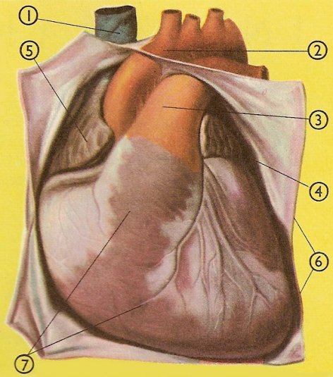
The heart and pericardium. 1. Superior vena cava. 2. Aorta. 3. Pulmonary artery. 4. Left atrium. 5. Right atrium. 6. Parietal pericardium. 7. Heart covered by the visceral pericardium.
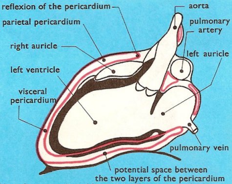
How the pericardium (in red) surrounds the heart.
The pericardium is the serous membrane (covering membrane) which, in humans and other vertebrates, forms the wall of the pericardial cavity and holds the heart in place. It is made of two thin layers of tissue.
Visceral pericardium (epicardium)
Immediately surrounding the heart, and attached to it, is the visceral pericardium, or epicardium. The heart can slide easily on the visceral pericardium, thus allowing it to beat freely. The visceral pericardium has an external layer of flat mesothelial cells, which lies on a stroma of fibrocollagenous support tissue. This support tissue contains elastic fibers, as well as the large arteries supplying blood to the heart wall, and the larger venous tributaries carrying blood from the heart wall.
Parietal pericardium
The outer layer of the pericardium, called the parietal pericardium, consists of an outer layer of strong, thick connective tissue (called the fibrous pericardium) and an inner serous layer (the serous pericardium). The fibrous layer of the parietal pericardium is attached to the diaphragm and fuses with the outer wall of the great blood vessels entering and leaving the heart. Thus, the parietal pericardium forms a strong protective sac for the heart and serves also to anchor it within the mediastinum. The serous layer of the parietal pericardium, composed largely of mesothelium together with a little connective tissue, forms a simple squamous epithelium and secretes a small amount of fluid (normally about 25 to 35 ml), which keeps the two layers of pericardium from rubbing against each other and causing friction during the heart's muscle contractions.
At the top of the heart the visceral layer folds over to join the parietal layer. This fold is called the reflexion of the pericardium.
A way to envisage the pericardium
Imagine that you have a football bladder filled with oil. The neck is untied and you press your fist downwards on to the bladder so that the oil runs out. When all but the last few drops have flowed away your fist will be surrounded on all sides and underneath by two layers of rubber which will slide easily on each other because of the small amount of oil that is left.
Now, if you pretend that your fist is your heart, the two layers of the football bladder will be like the two layers of the pericardium which surround it. At the top, your wrist will represent the large blood vessels which carry blood to and from the heart. The few drops of oil in the bladder will be the fluid between the two layers of pericardium which allows the layers to slide easily on each other. Although the outer layer of the pericardium is attached to the nearby tissues, the inner layer attached to the heart can slide on it quite easily, and so allow the heart to beat freely.
Pericardial cavity
The pericardial cavity, also called the pericardial sac, is the body cavity within which the heart lies. In vertebrates, it is a coelomic space, separated from the perivisceral part of the coelum (incompletely in elasmobranchs). In arthropods and molluscs, it is a hemocoelic space supplying blood to the heart.

