
skeletal muscle
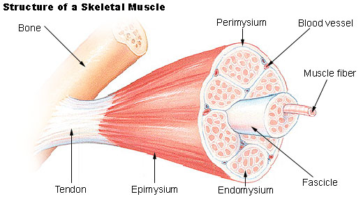
Figure 1. Structure of skeletal muscle.
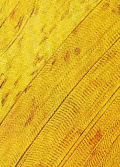
Figure 2. Skeletal, or voluntary, muscle consists of bundles of fibers. These are divided into bands, which appear as dark lines under a microscope. They contain actin and myosin, two important contractile proteins.
Skeletal muscle, also known as striated muscle or striped muscle, is a type of muscle, attached to bones, that is responsible for skeletal movements. The peripheral portion of the central nervous system (CNS) controls the skeletal muscles. Thus, these muscles are under conscious, or voluntary, control. The basic unit is the muscle fiber with many nuclei. Skeletal muscle fibers are striated (having transverse streaks) or striped and each acts independently of neighboring muscle fibers. Skeletal muscle makes up the bulk of muscles in the body. See also skeletal muscle groups.
A whole skeletal muscle is considered an organ of the muscular system. Each organ or muscle consists of skeletal muscle tissue, connective tissue, nerve tissue, and blood or vascular tissue.
Skeletal muscles vary considerably in size, shape, and arrangement of fibers. They range from extremely tiny strands such as the stapedium muscle of the middle ear to large masses such as the muscles of the thigh. Some skeletal muscles are broad in shape and some narrow. In some muscles the fibers are parallel to the long axis of the muscle, in some they converge to a narrow attachment, and in some they are oblique.
Each skeletal muscle fiber is a single cylindrical muscle cell. An individual skeletal muscle may be made up of hundreds, or even thousands, of muscle fibers bundled together and wrapped in a connective tissue covering. Each muscle is surrounded by a connective tissue sheath called the epimysium. Fascia, connective tissue outside the epimysium, surrounds and separates the muscles. Portions of the epimysium project inward to divide the muscle into compartments. Each compartment contains a bundle of muscle fibers. Each bundle of muscle fiber is called a fasciculus and is surrounded by a layer of connective tissue called the perimysium.
Skeletal muscle cells (fibers), like other body cells, are soft and fragile. The connective tissue covering furnish support and protection for the delicate cells and allow them to withstand the forces of contraction. The coverings also provide pathways for the passage of blood vessels and nerves.
Commonly, the epimysium, perimysium, and endomysium extend beyond the fleshy part of the muscle, the belly or gaster, to form a thick ropelike tendon or a broad, flat sheet-like aponeuroses (a thin but strong sheet of fibrous tissue). The tendon and aponeurosis form indirect attachments from muscles to the periosteum of bones or to the connective tissue of other muscles. Typically a muscle spans a joint and is attached to bones by tendons at both ends. One of the bones remains relatively fixed or stable while the other end moves as a result of muscle contraction.
Skeletal muscles have an abundant supply of blood vessels and nerves. This is directly related to the primary function of skeletal muscle, contraction. Before a skeletal muscle fiber can contract, it has to receive an impulse from a nerve cell (see muscle contraction). Generally, an artery and at least one vein accompany each nerve that penetrates the epimysium of a skeletal muscle. Branches of the nerve and blood vessels follow the connective tissue components of the muscle of a nerve cell and with one or more minute blood vessels called capillaries.
Origin and insertion of skeletal muscles
A skeletal muscle has two or more attachments. The attachment that moves the least is called the origin, and that which moves the most, the insertion. Under varying circumstances the degree of mobility of the attachments may be reversed, and therefore the terms origin and insertion are interchangeable.
Types of skeletal muscles
The individual fibers of a muscle are arranged either parallel or oblique to the long axis of the muscle. Since a muscle shortens by one-third to one-half its resting length when it contracts, then it follows that muscles whose fibers run parallel to the line of pull will bring about a greater degree of movement as compared with those whose fibers run obliquely. Examples of muscles with parallel arranged fibers are the sternomastoid, the rectus abdominus, and the sartorius.
Muscles who fibers run obliquely to the line of pull are referred to as pennate muscles (they resemble a feather). A unipennate muscle is one in which the tendon lies along one side of the muscle and the muscle fibers pass obliquely to it (e.g., extensor digitorum longus). A bipennate muscle is one in which the tendon lies in the center of the muscle and the muscle fibers pass to it from two sides (e.g., rectus femoris). A multipennate muscle (1) may be arranged as a series of bipennate muscles lying alongside one another (e.g., the acromial fibers of the deltoid) or (2) may have the tendon lying within its center and the muscle fibers passing to it from all sides, converging as they go (e.g., tibialis anterior).
For a given volume of muscle substance, pennate muscles have many more fibers as compared with muscles with parallel arranged fibers, and they are therefore more powerful; in other words, range of movement has been sacrificed to strength.
A-band and I-band
Two kinds of band are visible under certain conditions in a skeletal muscle fiber: A-band and I-band; alternation of the two kinds produces the cross-striations on skeletal muscle. The A-(anisotropic) band, unlike the I-(isotropic) band, contains myosin filaments.

