
cervical plexus
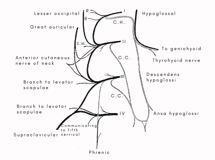
Cervical plexus and ansa hypoglossi. I, II, III, IV - anterior primary rami of the upper four cervical nerves. R. branches to rectiand longus capitis. S.M. branch to sternomastoid. C.C. roots of descendens cervicalis. C.H. communicating to hypoglossal.
The cervical plexus is formed by the anterior primary rami of the upper four cervical nerves. It is situated in the upper part of the neck opposite a line drawn down the side of the neck from the root of the auricle to the level of the upper border of the thyroid cartilage. It lies behind the internal jugular vein and the deep fascia, on the front of the transverse process of the atlas and on the scalenus medius muscle.
The four cervical nerves concerned in the formation of the plexus appear above the corresponding vertebrae, and they increase in thickness from above downward.
The anterior primary ramus of the first cervical appears at the medial margin of the rectus lateralis, descends over the front of the transverse process of the atlas and joins the second. The combined first and second pass downward and laterally to join the third on the front of the scalenus medius; and a communicating loop connects the third with the fourth. A slender branch that descends from the fourth to the fifth connects the cervical plexus with the brachial.
Branches of the cervical plexus
The branches are communicating, cutaneous, and muscular.
The communicating branches are: (1) The gray rami communicantes which the anterior primary rami of the upper four cervical nerves receive from the superior cervical ganglion. (2) A communicating branch from the first cervical to the hypoglossal. (3) Communicating branches from the second, third, and fourth to the accessory.
The cutaneous branches are the lesser occipital, great auricular, anterior cutaneous, and supraclavicular.
The muscular branches are the phrenic, the descendens cervicalis, and numerous branches which are distributed to the rectus capitis lateralis, the prevertebral, scalene, and inter-transverse muscles, the levator scapulae, the sternomastoid, and the trapezius. The phrenic nerve is the most important branch of the plexus since it is the nerve that supplies the chief muscle of respiration – the diaphragm.
Phrenic nerve
The nerve of the diaphragm arises chiefly from the fourth cervical nerve, and additional roots spring from the third and fifth. It begins in the scalenus medius at the lateral margin of the scalenus anterior, under cover of the sternomastoid, at the level of the upper border of the thyroid cartilage. It passes on to the scalenus anterior at once, and descends over the front of it between the muscular substance and the fascial covering – crossing the muscle from lateral to medial border. At the root of the neck, the right nerve passes off the scalenus anterior on to the pleura behind the innominate vein, and enters the thorax. The left nerve leaves the scalenus anterior at a higher level and descends in front of the first part of the subclavian artery into the thorax.
As the nerve lies in the scalenus anterior, it is under cover of the sternomastoid, at the lateral side of the internal jugular vein, and is overlapped by the vein. It is crossed by the inferior belly of the omohyoid, and branches of the thyrocervical trunk; the left nerve is crossed also by the thoracic duct. As the right nerve lies behind the innominate vein, it crosses the internal mammary artery from lateral to medial side, usually in front of the artery.
The root from the fifth cervical nerve usually joins the phrenic trunk on the surface of the scalenus anterior, but it may descend into the thorax before it effects a junction.

