duodenum

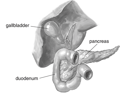
Duodenum, gallbladder, and pancreas. Image source: NIH.
The duodenum is the first and shortest part of the small intestine, being only about 10 inches (25 centimeters) long; it connects the stomach to the jejunum. It describes a C-shaped curve whose concavity is directed toward the left and upward and is occupied by the head of the pancreas. Owing to the C-shape of its curve, its beginning and end are not far apart. and
The duodenum receives chyme from the stomach through the pyloric sphincter. It also receives fluids from the pancreas and gallbladder via the common bile duct. The duodenum neutralizes the acidic chyme and mixes it with pancreatic, biliary, and intestinal secretions before passing it on to the jejunum.
The first inch (2.5 centimeter) of the duodenum resembles the stomach in that it is covered on its anterior and posterior surfaces with peritoneum and has the lesser omentum attached to its upper border and the greater omentum attached to its lower border; the lesser sac lies behind this short segment. The remainder of the duodenum is retroperitoneal, being only partially covered by peritoneum.
The duodenum is situated in the epigastric and umbilical regions, on the posterior wall of the abdomen above the level of the umbilicus, and almost wholly in the right half of the abdomen. All its parts do not lie in the same plane, because it is molded on the right side and the front of the median longitudinal elevation made by the spinal column and psoas muscle. The descending portion or right part of the C is thus much farther back than the rest of it. The principal changes of direction in the C-shaped are made use of to divide it into four parts for convenience of description.
First part of the duodenum
The first part is 2 inches (5 centimeters) long. It begins at the pylorus, in the transpyloric plane, about an inch to the right of the median plane, and passes sideward and backward and slightly upward in close relation with the liver, and ends at the neck of the gallbladder by bending sharply to become the second part. The first inch which appears in radiographs as the "duodenal cap", is clothed on the front and the back with the peritoneum continued on to it from the stomach. This portion is connected therefore with the omenta above and below; and, posteriorly, it is separated by the lesser sac of the peritoneum from the neck of the pancreas; anteriorly, it is related to the quadrate lobe of the liver. The second inch is clothed with peritoneum only above and in front, where it is related to the liver and the neck of the gallbladder. Inferiorly, it is related directly to the head of the pancreas. Posteriorly, it is directly related to the gastroduodenal artery, the bile duct, the portal vein, and a small portion of the neck of the pancreas; and these structures separate it from the inferior vena cava. It should be noted that, owing to its backward direction, these structure are medial to it rather than behind it.
The first inch, owing to its peritoneal connections, is free to move, and moves with the stomach. The second inch, like the other parts of the duodenum, is fairly firmly fixed by areolar tissue to the structures behind it.
Second part of the duodenum
The second part is 3 inches (7.5 centimeters) long. It descends to the level of the third lumbar vertebra and then bends at a right angle to become the third part. Anteriorly, it is crossed by the first part of the transverse colon, which covers most of it, and is overhung by the liver and the gallbladder; above the transverse colon, it is more directly related to the liver and the gallbladder; and its lowest part is covered by a loop of the jejunum. Posteriorly, it rests on the medial part of the right kidney and on the psoas major, being partly separated from the muscle by the renal vessels and the ureter. Laterally, it is related merely to the fat on the kidney. Medially, it is closely applied to the head of the pancreas. The bile duct and the pancreatic duct enter it together on its posteromedial surface a little below its middle.
The second part of the duodenum has a very incomplete covering of peritoneum – only on its parts of the anterior surface that are above and below the transverse colon, because the colon lifts the peritoneum off the greater part of it.
Third part of the duodenum
The third part is nearly 4 inches (10 centimeters). It begins on the right psoas major at the level of the third lumbar vertebra and passes nearly horizontally toward the left across the inferior vena cava and the aorta, and then bends upward to become the fourth part. The anterior and inferior surfaces are clothed with peritoneum except at its end where it is crossed by the superior mesenteric vessels and the root of the mesentery. On the right side of the mesentery it is covered by loops of the jejunum, which separate it from the transverse colon. Superiorly, it is closely applied to the head of the pancreas. Posteriorly, it rests on the right psoas, the inferior vena cava and the aorta, with the ureter, the testicular (or ovarian artery), and the inferior mesenteric artery intervening.
Fourth part of the duodenum
The fourth part is the shortest part – little more than an inch (2.5 centimeter) in length. It curves upward along the left side of the aorta and the head of the pancreas on to the left psoas muscle, and ends about an inch to the left of the median plane, at the level of the second lumbar vertebra, by bending sharply forward to form the duodenojejunal flexure, where it is continuous with the jejunum. On the front and left side it is covered with peritoneum and related to the jejunum. Behind, it is related to the left sympathetic trunk and the testicular (or ovarian) artery.
Variations in form and position of the duodenum
The curve of the duodenum varies with the position of the third part. Usually the third part is nearly horizontal and the fourth part nearly vertical; but the third part may incline upward as it passes toward the left, and lie in line with the fourth part.
There are, in addition, considerable variations not only in the position of the first part owing to its mobility, but also in the position of the whole duodenum in relation to the spinal column.
Vessels and nerves of the duodenum
The arteries are small branches from the hepatic, right gastric, pancreatico-duodenal, and right colic; the area of the mucous coat supplied by the duodenal branch of the hepatic artery is specially liable to the formation of a duodenal ulcer, and this is thought to be related to the fact that the artery has poor anastomoses with its neighbors. The nerves are derived from the celiac and superior mesenteric plexuses, and accompany the arteries. Thje lymph vessels end in glands that lie between the duodenum and the head of the pancreas, from where the lymph is carried to glands near the origins of the celiac and superior mesenteric arteries.


