
heart surgery
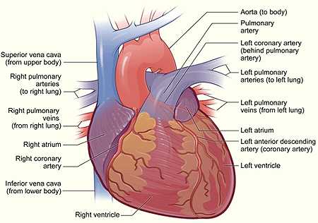
Exterior of the heart.
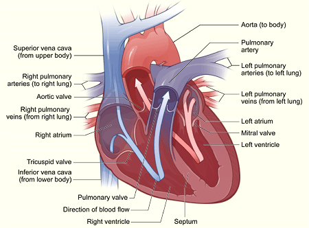
Interior of the heart.
Heart-lung bypass machine.
Heart surgery is done to correct problems with the heart. More than half a million heart surgeries are done each year in the United States alone for a variety of heart problems.
Heart surgery is used to correct heart problems in both children and adults. This article discusses types of heart surgery for adults. For more information about heart surgeries for children, see the articles on congenital heart defects, holes in the heart, and tetralogy of Fallot.
Overview
The most common type of heart surgery for adults is coronary artery bypass grafting (CABG). During CABG, surgeons use healthy arteries or veins taken from another part of the body to bypass (i.e., go around) blocked arteries. CABG relieves chest pain and reduces the risk of heart attack.
Heart surgery also is done to:
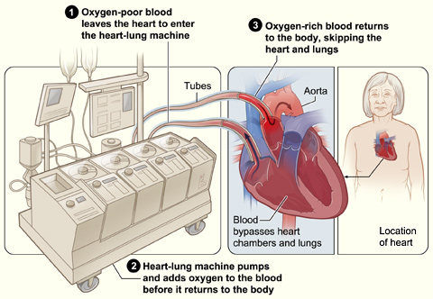
Traditional heart surgery, often called "open heart surgery," is done by opening the chest wall to operate on the heart. Almost always, the chest is opened by cutting through the patient's sternum (breastbone). Once the heart is exposed, the patient is connected to a heart-lung bypass machine, which takes over the pumping action of the heart. This allows surgeons to operate on the heart while it is still.
In recent years, new ways of doing heart surgery have been developed. One of these, called off-pump or beating heart surgery, is like traditional open-heart surgery but does not use a heart-lung bypass machine.
Minimally invasive heart surgery uses smaller incisions than traditional open-heart surgery. Some types of minimally invasive heart surgery use a heart-lung bypass machine and others don't.
These new methods may reduce risks and speed up recovery time. Studies are under way to compare these new types of heart surgery to traditional open-heart surgery. The results of these studies will help doctors decide the best procedure to use for each patient.
Outlook
The results of heart surgery in adults are often excellent. For very ill people with severe heart problems, heart surgery can reduce symptoms, improve quality of life, and increase lifespan.
Types of heart surgery
Different types of heart surgery are used to fix different heart problems.
Coronary artery bypass grafting
Coronary artery bypass grafting (CABG) is the most common type of heart surgery. More than half a million of these surgeries are done each year in the United States. CABG improves blood flow to the heart, and is used for people with severe coronary artery disease (CAD).
In CAD, a fatty material called plaque builds up inside the coronary arteries, narrowing the arteries and limiting blood flow to the heart muscle. CAD can cause angina (chest pain or discomfort), shortness of breath, and even lead to a heart attack.
During CABG, a surgeon takes a vein or an artery from the patient's chest, leg, or another part of the body and connects, or grafts, it to the blocked artery. The grafted artery bypasses the blockage, allowing oxygen-rich blood to reach the heart muscle. Surgeons can bypass as many as four blocked coronary arteries during one surgery.
Sometimes a patient has the choice between CABG and angioplasty to treat CAD. A doctor can be consulted about these different treatments.
Transmyocardial laser revascularization
Transmyocardial laser revascularization, or TLR, is a surgery used to treat angina when no other treatments work. For example, if a patient has already had one CABG procedure and can't have another one, TLR may be an option. This type of heart surgery is not common.
During TLR, the surgeon uses lasers to make channels in the heart muscle. These channels allow oxygen-rich blood to flow from a heart chamber directly into the heart muscle.
Valve repair or replacement
For the heart to work properly, blood must flow in only one direction. The heart's valves make this possible. Healthy valves open and close in a precise way as the heart pumps blood.
Each valve has a set of flaps called leaflet, which open to allow blood to pass from the heart chambers into the arteries. The leaflets then close tightly to stop blood from flowing back into the chambers.
Heart surgery is done to fix leaflets that don't open as wide as they should. This can happen when they become thick or stiff or fuse together. As a result, not enough blood flows through the valve into the artery.
Heart surgery is also done to fix leaflets that don't close tightly. This means blood can leak backward into the chambers, rather than only moving forward into the artery as it should.
To correct these problems, surgeons either repair the valve or replace it. Replacement valves are taken from animals, made from human tissue, or made from artificial substances.
Arrhythmia treatment
An arrhythmia is a problem with the speed or rhythm of the heartbeat. During an arrhythmia, the heart can beat too fast, too slow, or with an irregular rhythm.
Most arrhythmias are harmless, but some can be serious or even life-threatening. When the heart rate is abnormal, the heart may not be able to pump enough blood to the body. Lack of blood flow can damage the brain, heart, and other organs.
Arrhythmias are usually treated first with medicine. If medicines don't work well enough, a patient may need surgery, involving, for example, the insertion of a pacemaker or an implantable cardioverter defibrillator (ICD).
A pacemaker is a small device that's placed under the skin of the chest or abdomen. Wires lead from the pacemaker to the heart's chambers. The pacemaker sends electrical signals through the wires to control the speed of the heartbeat. Most pacemakers have a sensor that activates the device only when the heartbeat is abnormal.
An ICD is another small device that's placed in the chest or abdomen. This device also is connected to the heart with wires. It checks the heartbeat for dangerous arrhythmias and, if it senses one, sends a small electric shock to the heart to restore a normal heartbeat.
Another type of surgery for arrhythmia is known as maze surgery. In this operation, the surgeon makes new paths (a maze) for the heart's electrical signals to travel through. This type of surgery is used to treat atrial fibrillation, the most common type of serious arrhythmia.
Aneurysm repair
An aneurysm is an abnormal bulge or ballooning in the wall of an artery or the heart muscle. This bulge happens when the wall weakens. Pressure from blood moving through the artery or heart causes the weak area to bulge out. Over time an aneurysm can grow and eventually burst, causing dangerous, often fatal bleeding inside the body.
Aneurysms in the heart most often occur in the heart's lower left chamber, and can develop after a heart attack.
Repairing an aneurysm involves surgery to replace the weak section of the artery or heart wall with a patch or graft.
Ventricular assist devices
Ventricular assist devices (VADs) are mechanical pumps that support the heart or take over the heart's pumping action. VADs are used when the heart can't pump enough blood to support the body.
A patient may need a VAD if he or she has heart failure or is waiting for a heart transplant. A VAD can be used for a short time or for months or years, depending on the individual's situation.
Heart transplant
A heart transplant is surgery in which a diseased heart is replaced with a healthy heart from a deceased donor. Heart transplants are done on patients whose hearts are so damaged or weak that they can't pump enough blood to meet the body's needs.
This type of surgery is a life-saving measure that's used when medical treatment and less drastic surgery have failed.
Because donor hearts are in short supply, patients who need a heart transplant go through a careful selection process. They need to be sick enough to need a new heart, yet healthy enough to receive it.
Patients on the waiting list for a donor heart receive ongoing treatment for heart failure and other medical conditions. VADs may be used to treat these patients.
Surgical approaches
In recent years, new ways of doing heart surgery have been developed. Depending on a patient's heart problem, general health, and other factors, he or she can now have open-heart surgery or minimally invasive heart surgery.
Open-heart surgery
Open-heart surgery is any kind of surgery where the chest wall is opened and surgeons operate on the heart. "Open" refers to the chest, not the heart. Depending on the type of surgery, the heart may be opened too.
Open-heart surgery is used to bypass blocked arteries in the heart, repair or replace heart valves, fix atrial fibrillation, and transplant hearts.
In recent years, more surgeons have started to use off-pump, or beating heart, surgery to do CABG. This approach is like traditional open-heart surgery, but surgeons don't use a heart-lung bypass machine.
Off-pump heart surgery may reduce complications that can occur when a heart-lung bypass machine is used. It also may speed up recovery time.
Off-pump heart surgery isn't right for all patients. Doctors decide on a case by case basis whether this type of surgery is appropriate. He or she will carefully considers a patient's particular heart problem, age, overall health, and other factors that may affect the surgery.
Minimally invasive heart surgery
For minimally invasive heart surgery, a surgeon doesn't make a large incision (cut) down the center of the chest to open the rib cage. Instead, he or she makes small incisions in the side of the chest between the ribs.
A heart-lung bypass machine is used in some types of minimally invasive heart surgery, but not others.
This newer heart surgery is used for some CABG and maze procedures. It's also used to repair or replace heart valves and insert pacemakers.
One type of minimally invasive heart surgery that's still being developed is robotic-assisted surgery. For this surgery, a surgeon uses a computer to control surgical tools on thin robotic arms. The tools are inserted through small incisions in the chest. This allows surgeons to perform complex and highly precise surgery. The surgeon is always in total control of the robotic arms; they don't move on their own.
Benefits of minimally invasive heart surgery compared to open-heart surgery include smaller incisions and scars, lower risk of infection, less pain, a shorter hospital stay, and a faster recovery.
Who needs heart surgery?
Heart surgery is used to treat people who have severe heart diseases and conditions. If other treatments, such as lifestyle changes, medicines, and medical procedures, haven't worked or can't be used, heart surgery may be an option.
Heart surgery is used to treat heart failure and coronary artery disease. It's also used to fix heart valves that don't work right, to regulate heart rhythms, and to replace a damaged heart with a healthy one.
Specialists involved
The primary care doctor, a cardiologist, and a cardiothoracic surgeon will decide whether a particular patient needs heart surgery. A cardiologist specializes in treating heart problems. A cardiothoracic surgeon specializes in surgery on the heart and lungs.
These doctors will talk with a patient and do tests to learn about his or her general health and heart problem. They'll discuss test results with the patient, and will help him or her make decisions about the surgery.
Medical evaluation
A doctor will talk with a patient about:
The doctors also may do blood tests, such as a complete blood count, a cholesterol test, and other tests as needed.
Diagnostic tests
Medical tests are done to find out more about the heart problem and a patient's general health. This helps a doctor decide whether the patient needs heart surgery, what type of surgery is needed, and when to do it.
EKG (electrocardiogram)
An EKG is a simple and painless test that records the electrical activity of the heart. This test is used to help detect and locate the source of heart problems.
A technician attaches sticky patches, called electrodes, to the skin of the chest, arms, and legs. The electrodes are attached with wires to a machine that records the heart's electrical signals.
An EKG shows how fast the heart is beating and whether its rhythm is steady or irregular. It also shows where in the heart the electrical activity starts, and whether it's traveling through the heart in a normal way.
Stress test
Some heart problems are easier to diagnose when the heart is working harder and beating faster than when it's at rest. During stress testing, the patient exercises (or is given medicine if the patient is unable to exercise) to make the heart work hard and beat fast.
During the stress test, blood pressure is checked and an EKG is done. Other heart tests also may be performed.
Echocardiography
Echocardiography is a painless, noninvasive test. "Noninvasive" means that no surgery is done and no instruments are inserted into the body.
This test uses sound waves to create a moving picture of the heart. Echocardiography provides information about the size and shape of the heart and how well the heart chambers and valves are working.
The test also can show areas of poor blood flow to the heart, areas of heart muscle that aren't contracting normally, and previous injury to the heart muscle caused by poor blood flow.
Coronary angiography
Coronary angiography uses a special dye to show the insides of the coronary arteries on X-ray pictures. An angiogram shows the location and severity of blockages in blood vessels.
To get the dye to the coronary arteries, a procedure called cardiac catheterization is used. Cardiologists usually do cardiac catheterizations in a hospital. The patient is awake during the procedure, and usually experiences little to no pain.
During this procedure, a catheter (a thin, flexible tube) is passed through an artery in the leg or arm and threaded to the heart. The dye is injected into the bloodstream through the tip of the catheter.
Aortography
Aortography is angiography applied to aorta. The aorta is the main artery that carries blood from the heart to the body. An aortogram may show the location and size of an aortic aneurysm and the arteries that are involved.
Chest X-ray
A chest X-ray provides a picture of the organs and structures inside the chest, including the heart, lungs, and blood vessels. This test gives a doctor information about the size and shape of the heart. A chest X-ray also shows the position and shape of the large arteries around the heart.
Cardiac computed tomography scan
A cardiac computed tomography (CT) scan provides computer-generated, X-ray images of the internal organs. A liquid dye that can be seen on an X-ray is injected into a vein in the arm. The dye outlines arteries and veins in the heart on the CT scan.
A cardiac CT scan can show whether plaque is narrowing the coronary arteries or whether the patient has an aneurysm. A CT scan also can find problems with heart function and heart valves.
Cardiac magnetic resonance imaging
Cardiac magnetic resonance imaging (MRI) is a safe and noninvasive test that uses magnets and radio waves to create images of the inside of the body.
Cardiac MRI uses a computer to create images of the heart as it's beating. The computer makes both still and moving pictures of the heart and major blood vessels.
Cardiac MRI shows the structure and function of the heart. This test is very accurate at finding aneurysms and determining their size and exact location.
Before heart surgery
There are many types of heart surgery. The type a person need depends on his or her situation. One person's experience before an operation can be very different from another's.
Some people carefully plan their surgeries with their doctors. They know exactly when and how it will happen. Other people need emergency heart surgery. Others are diagnosed with blocked coronary arteries and are admitted to the hospital right away for surgery as soon as possible.
If a patient is having a planned surgery, he or she may be admitted to the hospital the afternoon or morning before the surgery. Doctors and others on the health care team will meet with the patient to explain what will happen. They will give the patient instructions on how to prepare for the surgery.
He or she also may need to have some tests, such as an EKG (electrocardiogram), chest X-ray, or blood tests. An intravenous (IV) line will be placed in the arm to administer fluids and medicines. Hair near the incision site may be shaved. The patient's skin may be washed with special soap to reduce the risk of infection.
Just before the surgery, the patient will be moved to the operating room and given medicine so that he or she falls asleep and feels no pain during the surgery.
During heart surgery
Heart surgery is done in a hospital. A team of experts is involved. Cardiothoracic surgeons perform the surgery with a team of other doctors and nurses who assist.
The length of time for the surgery depends on the type of surgery. CABG, the most common type of heart surgery, usually takes 3 to 5 hours.
Traditional open-heart surgery
For this type of surgery, the patient is given medicine to make him or her fall asleep. A doctor checks the patient's heartbeat, blood pressure, oxygen levels, and breathing during the surgery. A breathing tube is placed in his or her lungs through the throat and connected to a ventilator (breathing machine).
A surgeon makes a 6- to 8-inch incision (cut) down the center of the chest wall. The chest bone is cut and the rib cage is opened so that the surgeon can get to the heart.
The patient is given medicine to thin the blood and keep it from clotting. A heart-lung bypass machine is connected to the heart. This machine takes over for the heart by replacing the heart's pumping action. A specialist oversees the machine. The bypass machine allows the surgeon to operate on a heart that isn't moving and full of blood.
The patient is given medicines to stop the heartbeat once he or she is connected to the heart-lung bypass machine. A pipe is placed in the heart to drain blood to the machine. The machine removes carbon dioxide (a waste product) from the blood, adds oxygen, and then pumps the blood back into the body. Tubes are inserted into the chest to drain fluid.
Once the bypass machine begins to work, the surgeon performs the surgery to repair the heart problem.
At the end of the surgery, the heart is restarted using mild electric shocks. The pipes and tubes are removed from your heart, and the heart-lung bypass machine is stopped. The patient is given medicine to allow his or her blood to clot again.
The chest bone is closed with wires. Stitches or staples are used to close the incision. The breathing tube is removed.
An advantage of traditional open-heart surgery is that it's easier for the surgeon to operate. This is very important for long and complex surgeries.
Off-pump heart surgery
This type of surgery is the same as traditional open-heart surgery, except the patient is not connected to a heart-lung bypass machine. Instead, the heart is steadied with a mechanical device while the surgeon works on it. The heart continues to pump blood to the body.
The advantages of off-pump heart surgery are:
Minimally invasive heart surgery
For this type of heart surgery, the surgeon makes small incisions in the side of the chest between the ribs. These incisions can be as small as 2 to 3 inches. Then the surgeon inserts surgical tools through these small incisions. A tool with a small video camera at the tip also is inserted through an incision. This allows the surgeon to see inside the body.
Some types of minimally invasive heart surgery use a heart-lung bypass machine; other types don't.
The advantages of minimally invasive heart surgery are:
Patients who don't need the heart-lung bypass machine aren't at risk for the complications that the machine may cause.
After heart surgery
Recovery in the hospital
Depending on the type of heart surgery, a patient may spend 1 day or more in the hospital's intensive care unit. Then he or she is moved to another part of hospital for several days before going home.
While a patient is in the hospital, doctors and nurses will closely watch his or her heart rate, blood pressure, breathing, vital signs, and incision site(s). He or she may have an intravenous (IV) needle inserted in an arm to give fluids until he or she is ready to drink unassisted.
The patient also may be given extra oxygen through a face mask or nasal prongs that fit just inside the nose. These pieces of equipment are removed when the patient no longer needs them.
Recovery at home
Each person responds differently to heart surgery. An individual's recovery at home also will depend on what kind of heart problem and surgery he or she has had. A doctor will give specific instructions about how to:
A patient will also get information about follow-up appointments, medicines, and situations when he or she should call the doctor right away.
After-effects of heart surgery are normal. They may include:
Other after-effects may include loss of appetite, difficulty sleeping, constipation, and mood swings and depression. After-effects gradually go away.
Recovery time varies with type of heart surgery. Full recovery from traditional open-heart CABG may take 6 to 12 weeks or more. Less recovery time is needed for off-pump heart surgery and minimally invasive heart surgery.
A patient's doctor will let him or her know when it is safe to go back to daily activities, such as working, driving, and physical activity.
Ongoing care
Care after surgery may include periodic checkups with a doctor. During these visits, a patient may have blood tests, an EKG (electrocardiogram), an echocardiography, or a stress test. These tests will show how the heart is working after the surgery.
A patient's doctor also may talk to him or her about lifestyle changes and medicines to help stay healthy. Lifestyle changes may include quitting smoking, making changes in the diet, getting regular physical activity, and lowering and managing stress.
A doctor may refer a patient to a cardiac rehabilitation (rehab) program. Cardiac rehab includes counseling, education, and exercise training to help recovery. The program also will help a patient learn how to make choices that can lower the risk for future heart problems.
Risks
Heart surgery has risks, even though its results often are excellent. Risks can be from the surgery itself or from the heart-lung bypass machine. They include:
In general, the risks of heart surgery are higher for people who:
The use of a heart-lung bypass machine increases the risk of blood clots forming in the blood vessels. Clots can travel to the brain or other parts of the body and block the flow of blood. This can cause stroke or other problems. Recent improvements in heart-lung bypass machines are helping to reduce the risk of blood clots forming.

